Micro-Ultrasound vs MRI Prostate Biopsy: 2026 Evidence
The OPTIMUM trial showed micro-ultrasound matches MRI for finding aggressive prostate cancer. That is not the same as saying it should replace your MRI, and the difference decides whether you get biopsied at all.

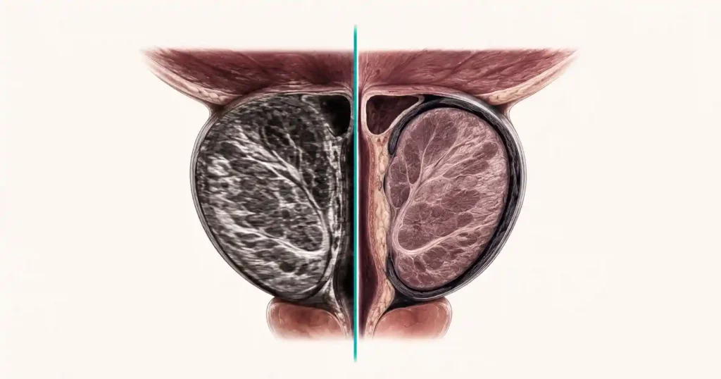
Micro-ultrasound vs MRI prostate biopsy is now a live question in my clinic, and it arrived faster than most men expect. Until 2025, the pathway was settled: raised PSA, then an MRI, then a targeted biopsy. Then the OPTIMUM randomized trial published in JAMA and showed that a 29 MHz ultrasound probe, used live in the biopsy room, found aggressive prostate cancer at least as often as an MRI-guided biopsy [1]. Health systems across the United States started installing the machines. Men started hearing the word in consultations. What almost nobody explains is that matching MRI on detection is not the same as replacing MRI in the pathway. The two do different jobs at different moments, and the moment matters more than the resolution. For the wider picture on screening and biopsy decisions, see our Prostate Health Hub.
Key Takeaways
- In the OPTIMUM trial, micro-ultrasound-guided biopsy found Grade Group 2 or higher cancer in 47.1% of men versus 42.6% with MRI fusion biopsy — a 3.52% difference that met the non-inferiority threshold.
- Micro-ultrasound is performed live by your urologist during the biopsy; MRI is read by a radiologist before you arrive. Only the MRI can tell you the biopsy is unnecessary.
- PRI-MUS scoring is highly sensitive but poorly specific — treating a score of 3 to 5 as suspicious catches 92% of significant cancers per lesion but is correct only about 55% of the times it fires.
- The AUA/SUO 2026 amendment upgraded MRI before initial biopsy to Grade A evidence and carries no equivalent recommendation for micro-ultrasound.
- Combining both modalities detected lesions that either one missed alone, which is why the strongest use case today is complementary, not replacement.
What Micro-Ultrasound Actually Is
The ultrasound probe used for a standard prostate biopsy runs at roughly 6 to 12 megahertz. That is enough to see the outline of the gland, the capsule and the needle, but not enough to see what the tissue inside is doing. Micro-ultrasound — commercially the ExactVu system — runs at 29 MHz, which lifts spatial resolution to around 70 microns, close to the scale at which tissue architecture becomes visible [5].
Think of it as the difference between a satellite photo that shows a city block and one that shows individual roof tiles. Prostate cancer disrupts the orderly ductal architecture of glandular tissue. At 29 MHz, that disruption becomes something the operator can see in real time rather than infer.
The trade-off is physical. Higher frequency buys resolution and loses penetration. The signal is excellent in the peripheral zone sitting closest to the probe, where most cancers arise, and degrades toward the front of the gland. Hold that fact — it comes back later.
What the OPTIMUM Trial Showed, and What Non-Inferior Really Means
OPTIMUM randomized 802 biopsy-naive men across 20 centers in 8 countries, of whom 678 went on to biopsy. Median age was 65 and median PSA was 6.9 ng/mL. Men were assigned to micro-ultrasound-guided biopsy, to a combined micro-ultrasound-then-MRI approach, or to conventional MRI fusion biopsy. Everyone also received systematic cores [1].
Grade Group 2 or higher cancer — the grade that generally warrants treatment rather than surveillance — was found in 47.1% of the micro-ultrasound group, 42.6% of the MRI fusion group and 46.9% of the combined group. The difference of 3.52% sat comfortably inside the pre-set 10% non-inferiority margin.
Here is where I ask patients to slow down. Non-inferior is a statistical verdict, not a clinical endorsement. The 95% confidence interval ran from minus 3.95% to plus 10.92%, which means the data are also compatible with micro-ultrasound being modestly worse. A 10% margin is generous. And every man in the trial received systematic cores alongside the targeted ones, which props up both arms — on targeted cores alone the rates were 38.0% and 34.1%, a difference that did not reach significance.
A 2026 systematic review of 15 studies and 2,512 patients reached the same place from a different direction: the pooled detection ratio between the two modalities was 1.06, with a confidence interval crossing 1.0 [3]. The honest summary is that these two tools perform similarly. Neither is beating the other.
Why MRI moved ahead of biopsy in the diagnostic order — and what that changed
Micro-Ultrasound vs MRI Prostate Biopsy: The Practical Differences
Detection rates are what trials measure. What changes your experience is everything around them — when the imaging happens, who interprets it, and what decision it can still influence by the time it exists.
| What it decides | Micro-ultrasound | MRI-first pathway |
|---|---|---|
| When the imaging happens | Live, in the biopsy room | A separate scan, days to weeks earlier |
| Who interprets it | The urologist holding the probe, in real time | A radiologist, reported before you attend |
| Scoring system | PRI-MUS 1 to 5 | PI-RADS 1 to 5 |
| GG2+ cancer found in OPTIMUM | 47.1% of men | 42.6% of men |
| Can it spare you the biopsy? | Not in practice — you are already prepped | Yes — a negative scan with low PSA density can defer it |
| Pacemaker, metal implant or claustrophobia | Usually no obstacle | Often rules the scan out |
| Guideline position in 2026 | No AUA recommendation statement yet | AUA/SUO 2026: may use before initial biopsy, Grade A |
Read the fifth row twice. It is the row that decides whether you have a needle in you at all.
Facing a prostate biopsy? Get the imaging and PSA-density checklist to take into your urology appointment
Enter your email below to receive Dr. Khalid’s complete PSA & Prostate Cancer Decision Guide as a free, printable PDF.
PRI-MUS Is Not PI-RADS, and the Gap Is Not Only Numerical
Both systems run 1 to 5 and both treat 3 and above as worth sampling, which invites men to assume they are interchangeable. They are not.
A secondary analysis of OPTIMUM scored 4,590 cores from 347 men. Treating PRI-MUS 3 to 5 as suspicious gave, at the lesion level, 92% sensitivity but only 34% specificity, with a positive predictive value of 55% [2]. In plain terms: a suspicious PRI-MUS reading misses very little, and is wrong roughly half the times it fires. It is a rule-out tool, not a rule-in tool.
The structural difference matters more than the statistics. A radiologist reads your MRI asynchronously — they can take time, compare sequences, and ask a colleague. PRI-MUS is scored in the moment, by the person already holding the biopsy gun, with you on the table. That removes a layer of second opinion from the pathway [5].
PI-RADS also carries formal backing from the American College of Radiology and is embedded in AUA guidance. PRI-MUS has no equivalent society adoption yet. If you want to see how those two numbers translate into a biopsy decision alongside your PSA density, our PI-RADS and PSA density biopsy decision tool walks through the same logic your urologist applies.
In My Practice
A man came to me last spring with a PSA of 7.4 ng/mL and an MRI report that read PI-RADS 2 — nothing to target. He had waited eleven weeks for that scan. His PSA density was 0.19 and his father had died of prostate cancer at 61, so I biopsied him regardless. We used micro-ultrasound to guide the cores, and it flagged an anterior area the MRI had called normal. It came back Gleason 4+3.
Neither imaging modality is a rule-out on its own; when PSA density and family history point one way and the scan points the other, the clinical picture wins.
The One Thing MRI Does That Micro-Ultrasound Cannot
The single largest benefit MRI brought to prostate care was never sharper targeting. It was avoided biopsies. Under European guidance, a man with a negative scan, a PSA density below 0.20, a normal digital rectal examination and no family history can reasonably defer biopsy and monitor his PSA instead. That decision is made weeks before he sets foot in the procedure room.
Micro-ultrasound structurally cannot deliver that. By the time the 29 MHz probe produces an image, you have had your bowel prep, your antibiotic and your local anesthetic, and the decision to biopsy has already been made. The imaging improves the aim; it does not reopen the question.
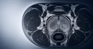
Two further things MRI does that a rectal probe cannot. It sees the anterior prostate well, exactly where 29 MHz penetration weakens, and anterior tumors are already the ones most often missed. And it stages — extraprostatic extension and seminal vesicle involvement change whether surgery spares the nerve bundles, and micro-ultrasound has no validated role there yet [6].
PSA density is the number that carries most of the weight in the avoided-biopsy decision, and most men never see it calculated. Run yours with our PSA density calculator before your next appointment so you know which side of 0.15 you sit on.
Who Should Ask About Micro-Ultrasound, and Who Should Not
Ask about it if any of these apply to you:
- You cannot have an MRI — a pacemaker, certain implants, severe claustrophobia, or a body habitus that defeats the scanner.
- Your MRI wait is measured in months and your PSA density is above 0.15, where delay carries real cost.
- You have had a negative biopsy but your PSA is still climbing, and repeating the same MRI is unlikely to produce new information.
- Your center offers it alongside MRI rather than instead of it — the combined arm found lesions each modality missed alone [3].
Do not push for it if your MRI is already booked within a few weeks and you have no contraindication. Asking to substitute micro-ultrasound in that situation trades away the possibility of avoiding the biopsy altogether — a worse deal than it sounds.
Two concrete things to say at your appointment. First: Is my biopsy performed on a 29 MHz micro-ultrasound platform, or conventional ultrasound? Ask the scheduling office in advance, not on the day. Second: Transrectal or transperineal? The AUA/SUO 2026 amendment accepts either route, but the transperineal approach avoids passing the needle through the rectal wall, which matters if you have had repeated courses of antibiotics or a resistant urinary infection in the past — see our guide to antibiotic resistance and recurrent infection for why that history changes the calculation [4].
When to Go to the ER After a Prostate Biopsy
Whichever imaging guided your cores, the post-biopsy risks are identical. Go to the emergency room the same day, not the next morning, if you develop any of these:
- A temperature of 38°C (100.4°F) or higher, especially with shaking chills — this can be sepsis and it moves fast.
- Complete inability to pass urine despite feeling full.
- Rectal or urinary bleeding that soaks through a pad, or passing clots you cannot pass through.
- Rising pelvic pain over 24 to 48 hours rather than settling.
Frequently Asked Questions
Does micro-ultrasound mean I can skip my prostate MRI?
Not yet. In OPTIMUM, micro-ultrasound matched MRI for finding aggressive cancer once the biopsy was already going ahead. What it cannot do is tell you the biopsy is unnecessary. A negative MRI with a low PSA density can defer a biopsy for months, whereas micro-ultrasound only reports once the probe is in place and you are prepped. That is the real micro-ultrasound vs MRI prostate biopsy difference.
My report says PRI-MUS 3. What does that actually mean?
PRI-MUS 3 is the threshold at which urologists start taking targeted cores. In the OPTIMUM secondary analysis, treating 3 to 5 as suspicious caught 92% of significant cancers per lesion but was correct only about 55% of the times it fired. So a 3 usually means cores were taken from that spot, not that cancer was found. It behaves much like an indeterminate PI-RADS 3 on MRI.
Is micro-ultrasound available where I live in the US?
Availability is patchy and center-dependent. The ExactVu system is FDA-cleared and installed in a growing number of academic centers and large private urology groups, but most community hospitals still run conventional 6 to 12 MHz ultrasound. Ask the scheduling office directly whether your biopsy is performed on a 29 MHz platform, and ask before your pre-biopsy appointment rather than on the day.
Does micro-ultrasound make the biopsy less painful or lower my infection risk?
No. Micro-ultrasound changes how targets are chosen, not how cores are taken. Discomfort and infection risk are driven by the route, the number of cores and the antibiotic prophylaxis, not by the imaging frequency. If the procedure itself is what worries you, our walkthrough of what a prostate biopsy actually feels like covers the parts men are rarely told about.
My MRI was clear but my PSA keeps climbing. Would micro-ultrasound help?
Possibly, as a second look. Both OPTIMUM and the 2026 meta-analysis found lesions that each modality missed alone, which is why the combined arm performed best on targeted cores. If your PSA density is above 0.15 and the MRI is negative, a micro-ultrasound-guided biopsy is a more useful next conversation than repeating the same scan and expecting a different answer.
References
- Kinnaird A, Luger F, Cash H, et al. Microultrasonography-guided vs MRI-guided biopsy for prostate cancer diagnosis: the OPTIMUM randomized clinical trial. JAMA. 2025;333(19):1679-1687. PubMed
- Lazarovich A, Guer M, Luger F, et al. External validation of the PRI-MUS scoring system: a secondary analysis of the OPTIMUM randomized controlled trial. J Urol. 2026;216(3):352-359. PubMed
- Besiroglu H, Erkoc M, Cakir SS, et al. Micro-ultrasound versus mpMRI for targeted prostate biopsy: a systematic review, meta-analysis, and clinical integration. Ther Adv Urol. 2026;18:17562872261476755. PubMed
- Lin DW, Carlsson S, Filson CP, et al. Updates to early detection of prostate cancer: AUA/SUO guideline (2026). J Urol. 2026. AUA
- DuBois J, Smani S, Golos A, et al. Micro-ultrasound in the detection of clinically significant prostate cancer: a comprehensive review and comparison with multiparametric MRI. Tomography. 2025;11(7):80. PubMed
- Ashrafi D, Klotz L. From detection to surveillance: the evolving role of micro-ultrasound in prostate cancer. Diagnostics (Basel). 2026;16(15):2364. PubMed
Dr. Muhammad Khalid
MBBS · FCPS (Urology) · MCPS (General Surgery) · CHPE · CRSM · IMC #539472
Specialist urologist with 11+ years of clinical experience across tertiary teaching hospitals. Trained at Lady Reading Hospital and Khyber Teaching Hospital, Peshawar. Author or co-author of five peer-reviewed international publications, including in Cureus and the World Journal of Surgery. Procedural expertise: URS, PCNL, RIRS, TURP, TURBT, and major open urological surgery. Full profile →
This article is for educational purposes only and does not constitute medical advice. Always consult your physician or urologist for diagnosis and treatment decisions specific to your condition.