Prostate Volume Formula: How Your Size Is Calculated
The prostate volume formula assumes your prostate is a perfect ellipsoid. It is not — and the gap between the number on your report and your actual gland can decide whether you are offered a biopsy.

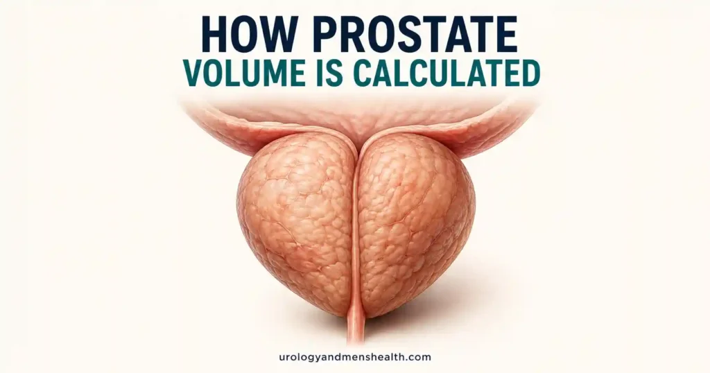
The prostate volume formula is simple arithmetic: length × width × height × 0.52. That one line of math produces the number written on your ultrasound or MRI report, and it carries more weight than most men realize. It influences whether finasteride is worth taking, which surgical options you will be offered, and whether your PSA result reads as reassuring or suspicious.
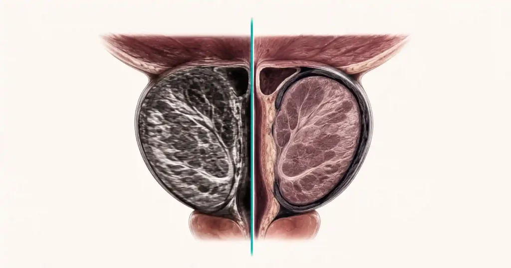
What rarely gets explained is that the formula treats your prostate as a perfect ellipsoid — a smooth, symmetrical egg. Real prostates are not. They have median lobes, asymmetric transition zones and irregular contours that three straight-line measurements cannot capture. The output is a number with a known error margin that shifts depending on who measured you and on which machine. For how size fits alongside PSA, symptoms and treatment, our Prostate Health Hub maps the whole pathway.
I go through this in clinic most weeks, usually with a man holding two reports that disagree and wanting to know which one is real. Here is how the number is produced, how far off it can be, and which decisions genuinely hinge on it.
Key Takeaways
- The prostate volume formula is length × width × height × 0.52 — the geometry of a perfect ellipsoid, which no real prostate is.
- Measured against removed glands, transrectal ultrasound underestimated true size by more than 30% in 55% of men, and overestimated it in only 6.4%.
- When seven radiologists measured the same MRI scans with the ellipsoid formula, their disagreement was enough to flip PSA density across the 0.15 biopsy threshold in 17.5% of patients.
- The two numbers that change management are 30 mL and 80 mL — below 30 mL a 5-alpha reductase inhibitor is unlikely to earn its place, and above 80 mL the surgical conversation changes entirely.
The Prostate Volume Formula: Three Numbers and One Constant
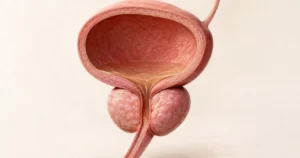
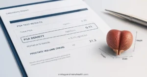
Every prostate volume on every report comes from the same three measurements. Length runs base to apex, taken on a sagittal image. Width is the side-to-side transverse diameter. Height is front to back, the anteroposterior diameter. Multiply the three, then multiply by 0.52.
The 0.52 is not a fudge factor someone settled on. It is π/6 rounded to two decimal places — the exact geometric constant for the volume of a prolate ellipsoid, the egg shape the formula assumes your gland to be.
A typical report might list 4.5 cm × 5.0 cm × 3.8 cm (about 1.8 × 2.0 × 1.5 inches). The arithmetic runs: 4.5 × 5.0 × 3.8 = 85.5, then 85.5 × 0.52 = 44.5 mL. That is the whole calculation.
Units cause needless confusion. Milliliters, cubic centimeters and cc are identical — 44.5 mL, 44.5 cm³ and 44.5 cc are the same gland. Grams differ only slightly: prostate tissue runs around 1.05 g per mL, so a 44.5 mL prostate weighs roughly 47 g. If your report quotes grams and a previous one quoted milliliters, that is not growth. If you want to check the math on your own report, our prostate volume calculator runs the same equation and grades where the result sits.
Where the Three Measurements Come From — and Why They Disagree
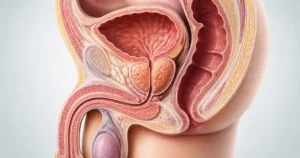
Transrectal ultrasound (TRUS) sits the probe a centimeter from the gland, which gives the clearest borders. Width and height are taken on the transverse view, length on the sagittal view. This is the method most volume figures in urology come from.
Transabdominal ultrasound images through a filled bladder from the lower abdomen. It is quick and needs no preparation beyond a full bladder, which is exactly its weakness: bladder filling, body habitus and probe angle all shift the borders, and the bladder base is easily included in the height measurement.
MRI allows two approaches. Radiologists can apply the same ellipsoid formula to T2 images, or they can perform planimetry — outlining the gland on every slice and summing the areas. Planimetry is the reference standard, takes far longer, and is why an MRI volume and an ultrasound volume are not interchangeable numbers.
A digital rectal exam sits outside all of this. My finger reaches the posterior surface only, so DRE systematically undercalls size, and a gland extending upward into the bladder can feel unremarkable.
The dimensions are not equally trustworthy. In the study that examined this most directly, comparing ultrasound measurements against the same glands after removal, length was accurate and width was the least reliable dimension — the opposite of what had been assumed[1]. Width is also the one being multiplied into every calculation.
Practical step: before starting finasteride or dutasteride, ask for a documented volume and the method that produced it, and ask that the repeat at twelve months uses the same method. The AUA notes that gland reduction on a 5-alpha reductase inhibitor is greater in larger prostates[4], and you cannot see that reduction if the measurement technique changed underneath you.
Enlarged prostate: what your size number means and what to ask for next
Enter your email below to receive Dr. Khalid’s complete BPH & Enlarged Prostate Guide as a free, printable PDF.
How Wrong Your Number Can Be
Three studies frame the problem, and they do not all point the same direction — which is itself the finding worth understanding.
Rodriguez and colleagues compared ultrasound volumes in 181 men against the weight of the glands after radical prostatectomy. Ultrasound underestimated by more than 30% in 55% of cases and overestimated by more than 10% in only 6.4%. More striking: when they fed the pathologists’ own measured dimensions into the formula, it still landed within 10% of true weight in just 26.5% of cases. The shape assumption, not the scanning, was the main source of error[1].
Haas and colleagues tested the formula on 3-Tesla MRI in 143 men against planimetry. Agreement was strong (r = 0.903) with a consistent lean toward underestimation — median 43.1 mL by formula against 48.1 mL by planimetry, roughly a tenth of the gland missing[2].
Hamzaoui and colleagues then had seven radiologists independently measure the same 40 MRI scans. Here the ellipsoid formula slightly overestimated against planimetry. The number that matters clinically is different: variation between raters was enough to move men across the PSA density threshold of 0.15 in 17.5% of cases using the ellipsoid formula, against 5% using planimetry[3].
Read together: the direction of the bias depends on what you compare against — a removed gland or an MRI outline — but the magnitude is consistent at roughly 10% to 20%, and a meaningful part of it comes down to who held the probe. Treat your volume as a band of about plus or minus 15%, not a fixed figure. That band matters most when the number is being divided into your PSA, which is covered in detail in how prostate volume and PSA density work together.
In My Practice
A man arrived with two reports six weeks apart — 52 mL from a transabdominal scan at a screening clinic, 41 mL by transrectal ultrasound at my hospital. He had spent those six weeks certain his prostate was growing fast enough to need surgery. Nothing had grown. His bladder was fuller for the first scan, and the abdominal view had swept part of the bladder base into the height measurement.
When two volumes disagree, the first question is which method produced each number, not which prostate changed.
What Your Prostate Volume Number Actually Changes
Volume is not a score of how bad things are. It is an eligibility variable — it determines which doors are open.
| Volume band | What it usually reflects | What it changes |
|---|---|---|
| Under 30 mL (about 1 fl oz) | Gland within the typical adult range; obstruction, if present, is often from a median lobe or bladder neck rather than bulk | A 5-alpha reductase inhibitor has little to work with; bladder function and outlet anatomy deserve the attention instead |
| 30 to 80 mL | Enlargement that crosses the thresholds used in guideline recommendations | AUA 2026 supports combining a 5-alpha reductase inhibitor with an alpha blocker above 30 mL; prostatic urethral lift is an option in the 30 to 80 mL range when no obstructing median lobe is present |
| Over 80 mL (about 2.7 fl oz) | Substantial enlargement, often with a long symptom history and higher residual volumes | AUA 2026 offers simple prostatectomy — open, laparoscopic or robotic — as an option at this size; enucleation techniques are usually part of the same discussion |
| Any band, with a raised PSA | Volume becomes the denominator of PSA density | Every error in the volume estimate is inherited by the density figure, which is why it is read alongside MRI findings rather than alone |
The 30 mL line comes from AUA 2026 Part II, which supports offering a 5-alpha reductase inhibitor alongside an alpha blocker when estimated volume exceeds 30 cc and/or PSA exceeds 1.5 ng/mL, at Moderate Recommendation, Evidence Level Grade B[4]. The 80 mL line comes from Part III, which offers simple prostatectomy for glands above that size, again Moderate Recommendation, Grade B, and confines prostatic urethral lift to the 30 to 80 cc range without an obstructing median lobe[5]. Those procedural options are unpacked further in the full guide to when an enlarged prostate needs surgery.
Notice what volume does not predict: how bothered you are. A 90 mL gland can produce a strong stream and an untroubled night, while a 28 mL gland with a median lobe sitting on the bladder neck can produce severe urgency and frequency. When symptoms are dominated by urgency, frequency and nocturia rather than a weak stream, the bladder is often the driver, and bladder training for an overactive bladder can do more than anything aimed at the prostate. Ask your urologist for a flow rate and a post-void residual alongside the volume — those describe the obstruction; volume only describes the anatomy.
Red Flags That Outrank Any Volume Number
No prostate size makes these safe to sit on, and no small prostate makes them harmless:
- Complete inability to pass urine with a painful, distended lower abdomen — go to the emergency room the same day for catheter drainage.
- Visible blood in the urine, with or without clots — request a urology referral within days for a urine test and imaging, whatever your prostate measures.
- Fever, shivering or back pain alongside urinary symptoms — same-day medical assessment; this is infection, not obstruction.
- New nausea, confusion or a sharp drop in urine output in a man with a longstanding poor stream — ask for kidney function bloods and a bladder scan urgently, as this pattern can signal back-pressure on the kidneys.
Frequently Asked Questions
Why does my prostate volume differ between my ultrasound and my MRI report?
Because those are two different measurements, not one measurement repeated. Transrectal ultrasound, an abdominal scan and MRI each define the gland borders slightly differently, and multiplying three diameters together amplifies small differences. A gap of 10 to 20 percent between two reports is common and rarely means anyone made a mistake. What matters is using the same method whenever you are tracking change over time.
Is a prostate volume of 40 mL big enough to explain my symptoms?
It can be, but size alone never settles it. Plenty of men with 40 mL glands have no symptoms at all, and plenty with 25 mL glands are badly obstructed by a small median lobe at the bladder neck. Your flow rate, post-void residual and symptom score describe the obstruction. Volume mainly tells your urologist which treatments you are eligible for.
Does the prostate volume formula work if my prostate has a median lobe?
Less well. A median lobe grows upward into the bladder, and that intravesical portion is often partly missed when the three diameters are taken, so the calculated figure understates the gland. This matters twice over, because a median lobe also rules out certain procedures. If your report mentions intravesical protrusion, ask whether that portion was included in the volume.
My report says grams instead of milliliters — is that the same number?
Close enough for any decision you will face. Prostate tissue is slightly denser than water, around 1.05 grams per milliliter, so a 40 mL gland weighs roughly 42 grams. Cubic centimeters, cc and mL are identical units with no conversion at all. The real difference between two reports comes from the measurement, not the unit.
How does my prostate volume change how my PSA is read?
Through PSA density, which is your PSA divided by your volume. A PSA of 6 in a 90 mL gland gives a density of 0.07; the same PSA in a 30 mL gland gives 0.20, and those two men receive very different biopsy advice. Because density inherits every error in the volume estimate, it is interpreted alongside MRI findings rather than alone. Our PSA density calculator works out where you sit.
Can I calculate my own prostate volume from the measurements in my report?
Yes, provided your report lists all three diameters in centimeters. Multiply length by width by height, then by 0.52 — that is the prostate volume formula radiologists use. Our prostate volume calculator does the arithmetic and grades the result. Treat the answer as a band of roughly plus or minus 15 percent rather than an exact figure.
References
- Rodriguez E Jr, Skarecky D, Narula N, Ahlering TE. Prostate volume estimation using the ellipsoid formula consistently underestimates actual gland size. Journal of Urology. 2008;179(2):501-503. PubMed
- Haas M, Günzel K, Miller K, Hamm B, Cash H, Asbach P. Is the ellipsoid formula the new standard for 3-Tesla MRI prostate volume calculation without endorectal coil? Urologia Internationalis. 2017;98(1):49-53. PubMed
- Hamzaoui D, Montagne S, Granger B, et al. Prostate volume prediction on MRI: tools, accuracy and variability. European Radiology. 2022;32(7):4931-4941. PubMed
- American Urological Association. Management of Lower Urinary Tract Symptoms Attributed to Benign Prostatic Hyperplasia: AUA Guideline (2026), Part II: Medical Management. Journal of Urology. 2026. AUA
- American Urological Association. Management of Lower Urinary Tract Symptoms Attributed to Benign Prostatic Hyperplasia: AUA Guideline (2026), Part III: Procedural/Surgical Management. Journal of Urology. 2026. PubMed
Dr. Muhammad Khalid
MBBS · FCPS (Urology) · MCPS (General Surgery) · CHPE · CRSM · IMC #539472
Specialist urologist with 11+ years of clinical experience across tertiary teaching hospitals. Trained at Lady Reading Hospital and Khyber Teaching Hospital, Peshawar. Author or co-author of five peer-reviewed international publications, including in Cureus and the World Journal of Surgery. Procedural expertise: URS, PCNL, RIRS, TURP, TURBT, and major open urological surgery. Full profile →
This article is for educational purposes only and does not constitute medical advice. Always consult your physician or urologist for diagnosis and treatment decisions specific to your condition.