
Kidney Stone Passage Calculator
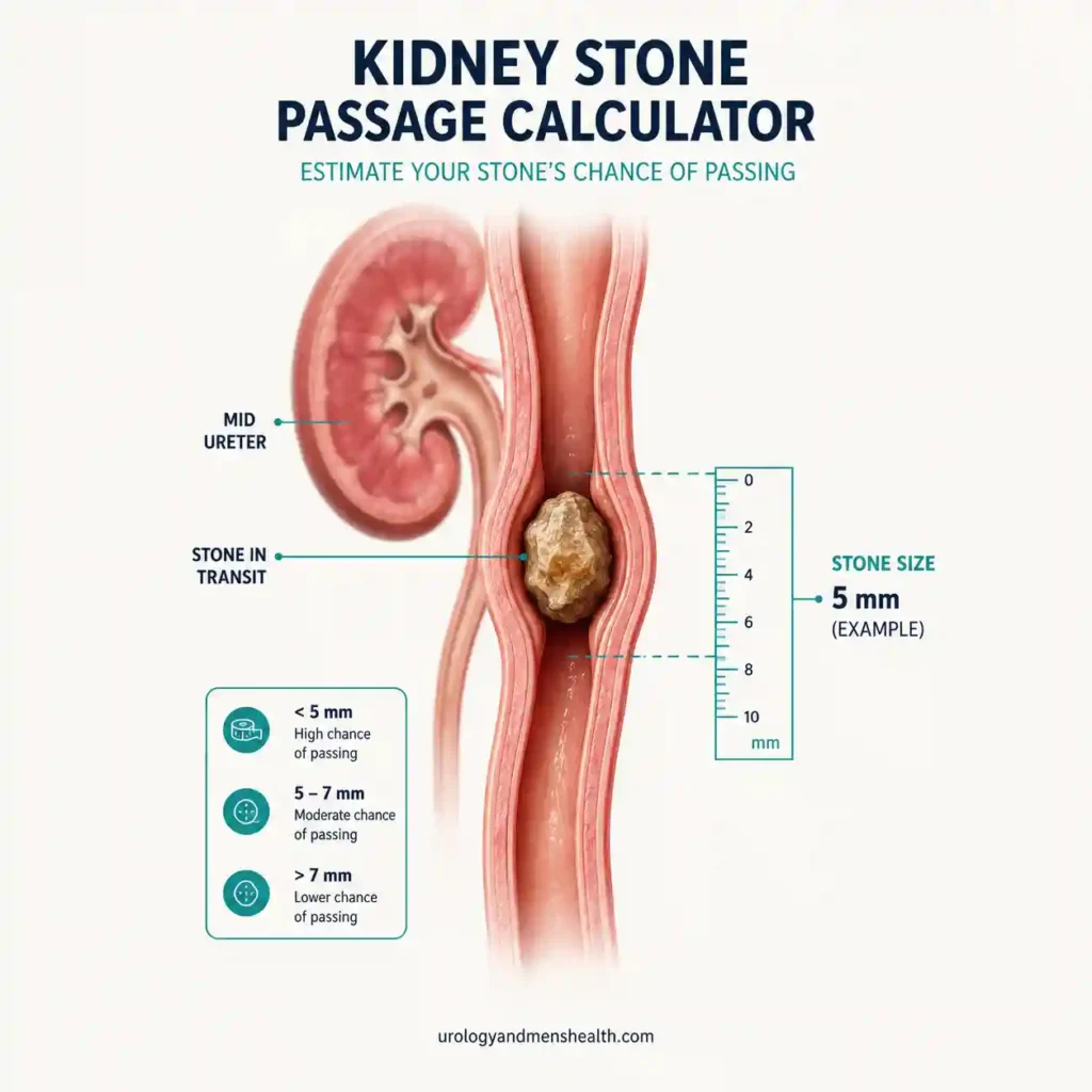
This kidney stone passage calculator estimates the chance your stone will pass on its own, using the two strongest predictors urologists rely on: its size in millimeters and its position in the urinary tract. A 3 mm stone near the bladder behaves very differently from an 8 mm stone still in the kidney. You get a probability, a clear next step, and a printable report to take to your kidney stone resources appointment.

The Tool
Related Kidney Tools
Full Clinical Guide
In This Guide:
- Size is the strongest predictor. Stones 4 mm or smaller pass on their own in roughly 80% of cases; the odds fall sharply once a stone is above 5 mm.
- Location matters as much as size. A stone low in the ureter has already cleared the hardest stretch and is far closer to passing than one still in the kidney or upper ureter.
- There is a time limit. Most stones that pass do so within about two to three weeks; waiting much longer risks infection or quiet kidney damage behind the blockage.
- Medication can shift the odds. Alpha-blockers such as tamsulosin relax the lower ureter and improve passage of distal stones up to 10 mm.
What This Tool Measures
This kidney stone passage calculator estimates the statistical likelihood that a stone leaves your body on its own, without surgery. It models the two predictors that decades of imaging studies have shown to matter most: the maximal diameter of the stone in millimeters, and where it currently sits in the collecting system or ureter. The prediction approach follows the validated size-and-location model published by Jendeberg and colleagues, who tracked 392 ureteral stones to passage or 26 weeks, and is aligned to the American Urological Association (AUA) and European Association of Urology (EAU) urolithiasis guidelines [1]. It tells you which way the odds lean — it does not replace a CT scan or a urologist’s judgment.
The Physiology of Stone Passage
To pass, a stone has to travel the length of the ureter — a muscular tube that is only a few millimeters wide at its tightest points. As the stone presses through, it stretches the ureteral wall and sets off the muscular spasm that produces the severe flank pain of renal colic. There are three natural narrow points where stones tend to lodge: the ureteropelvic junction where the kidney drains into the ureter, the spot where the ureter crosses the pelvic blood vessels mid-course, and the ureterovesical junction (UVJ) where it tunnels into the bladder wall.
Size sets the ceiling on what is physically possible. Spontaneous passage can be expected in up to 80% of stones no larger than 4 mm [2]. In the granular imaging data, passage falls to about 65% at 5 mm, around 33% at 6 mm, and under 10% once a stone reaches roughly 6.5 mm in width [1]. For stones wider than 7 mm, the EAU notes the chance of spontaneous passage is very low [2].
How to Interpret Your Result
Two people can sit in the same waiting room with stones that need opposite plans. A 3 mm stone sitting at the UVJ has already cleared almost the entire ureter and will very likely pass with fluids and time; an 8 mm stone still in the renal pelvis has the whole ureter ahead of it and is unlikely to pass at all. That is why this tool weighs location alongside size: among stones under 5 mm, the EAU reports an 89% passage rate in the distal ureter versus 71% in the upper ureter [2]. A High Likelihood result supports a trial of spontaneous passage with pain relief and hydration. A Moderate Likelihood result is the grey zone — passage is possible, but a firm two-to-four-week deadline should be agreed with your urologist. A Low Likelihood result means the dimensions make passage unlikely, and moving toward a procedure protects kidney function.
Related read: Kidney Stone Size Chart — When to Wait vs When to Book SurgeryWhat to Do With Your Result
If your probability is high, ask about medical expulsive therapy: the AUA recommends offering an alpha-blocker for roughly 30 days for distal ureteral stones up to 10 mm, with tamsulosin 0.4 mg once daily the usual choice — it relaxes the lower ureter and improves passage [3]. Strain your urine, keep any fragment for composition analysis, and stay consistently hydrated (around 2.5 liters / 85 fl oz of fluid a day). If your probability is moderate, treat the trial as time-limited and book repeat imaging at the deadline rather than relying on how you feel. If it is low, a planned procedure — ureteroscopy, shock wave lithotripsy, or PCNL for larger stones — is usually the most efficient route, and waiting mainly risks silent obstruction. Once this episode is settled, your long-term recurrence risk is the next thing worth checking. If you are unsure about your result, the PDF report this tool generates gives you a ready-made framework to bring to your next appointment.
Related read: How to Pass a Kidney Stone Fast — Evidence-Based ProtocolIn My Practice
The trap I warn every stone patient about is the day the pain stops. It feels like victory, and sometimes it is — the stone has dropped into the bladder. But just as often the stone has settled into a position where it no longer stretches the ureter while still blocking it. I have repeated scans on people who were certain they had passed their stone weeks earlier, only to find it sitting exactly where it was, the kidney quietly under pressure.
Pain is an unreliable witness. The only proof a stone has passed is catching it in a strainer, or a scan that shows it gone.
References
- Jendeberg J, et al. Size matters: the width and location of a ureteral stone accurately predict the chance of spontaneous passage. Eur Radiol, 2017. Prediction model from 392 ureteral stones (81% at 4 mm, 65% at 5 mm, 33% at 6 mm, 9% at ≥6.5 mm).
- European Association of Urology. EAU Guidelines on Urolithiasis. Up to 80% passage for stones ≤4 mm; distal <5 mm 89% vs upper 71%; very low passage above 7 mm; mean expulsion about 17 days.
- American Urological Association. Surgical Management of Kidney and Ureteral Stones: AUA Guideline (2025). MET with alpha-blockers for ~30 days for distal ureteral stones ≤10 mm (Strong Recommendation, Grade A).
- Preminger GM, et al. 2007 AUA/EAU Guideline for the Management of Ureteral Calculi. Meta-analysis: 68% passage for stones ≤5 mm, 47% for 5–10 mm.
Frequently Asked Questions
Does medication actually help a stone pass on its own?
Yes, for the right stones. Medical Expulsive Therapy uses an alpha-blocker, most often tamsulosin 0.4 mg, to relax the smooth muscle of the lower ureter so it dilates slightly and spasms less. Current AUA guidance supports offering it for about 30 days for distal ureteral stones up to 10 mm. It does little for very small stones, which pass anyway, or for large ones too wide for the ureter. See our evidence-based protocol for passing a stone faster.
Does drinking more water flush the stone out faster?
Not the way most people hope. Once a stone is lodged, forcing large volumes of water will not push it through, and if the blockage is complete it can briefly raise the pressure behind it and worsen pain. What steady hydration does do is keep urine flowing, reduce new crystal formation, and support flow around partial blockages. Aim for a consistent intake of around 2.5 liters (about 85 fl oz) a day rather than occasional large amounts. Our guide to hydration and kidney health covers the detail.
If the pain suddenly stops, does that mean the stone passed?
Not always, and this matters. Sudden relief often means the stone has dropped into the bladder, from where it passes within hours. But pain can also stop because the stone shifted into a position where it no longer stretches the ureter while still obstructing it — silent obstruction that can damage the kidney over weeks. The only proof is catching the stone in a strainer or a follow-up scan showing it gone. Our kidney stone size chart explains when imaging confirmation is needed.
How accurate is this kidney stone passage calculator, and can I rely on it?
It gives a statistical estimate from the two most reliable predictors in urology — stone size and location — drawing on AUA and EAU guideline data. No tool is exact for one person: prior stone-passing history, ureteral anatomy such as strictures, and stone texture all play a part. Treat it as an evidence-based screening aid, not a substitute for imaging or a urologist’s judgment. For the full clinical picture, read our complete guide to kidney stone symptoms and treatment.
How do I use this result at my doctor’s appointment?
Click the Download My Report (PDF) button below your result. It generates a printable two-page summary with your estimated passage probability, your stone’s size and location, and targeted questions to ask about conservative management versus surgery. Hand it over at the start of the consultation to anchor the discussion on your specific stone. Once the acute episode is settled, it is also worth checking your long-term odds with our 5-Year Kidney Stone Recurrence Risk Profiler.
Dr. Muhammad Khalid
MBBS · FCPS (Urology) · MCPS (General Surgery) · CHPE · CRSM · IMC #539472
Specialist urologist with 11+ years of clinical experience across tertiary teaching hospitals. Trained at Lady Reading Hospital and Khyber Teaching Hospital, Peshawar. Author or co-author of five peer-reviewed international publications, including in Cureus and the World Journal of Surgery. Procedural expertise: URS, PCNL, RIRS, TURP, TURBT, and major open urological surgery. Full profile →
This article is for educational purposes only and does not constitute medical advice. Always consult your physician or urologist for diagnosis and treatment decisions specific to your condition.