Blood in Urine in Men Over 60: Automatically High-Risk
The 2025 AUA amendment loosened the age bands for women and left one number untouched: 60. Blood in urine in men over 60 lands you in the high-risk group on age alone, before anyone counts a single red cell.

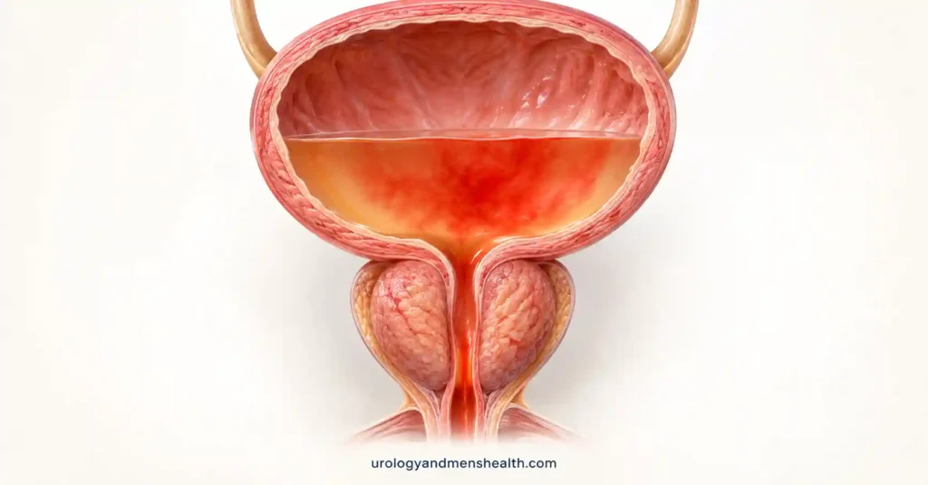
Blood in urine in men over 60 is not a scaled-up version of the same problem a 35-year-old has, and the workup that follows should not be the same either. That difference is written directly into the guideline. When the American Urological Association and SUFU amended their microhematuria guideline in 2025, they relaxed the age thresholds for women and left the male thresholds exactly where they were. A man aged 60 or older with confirmed microscopic blood in the urine sits in the high-risk group on age alone — before anyone asks about smoking, before anyone counts how many red cells appeared on the slide [1]. High-risk is not a mood or a warning label. It is a specific instruction: cystoscopy plus axial upper tract imaging. Not a repeat dipstick in six months. Not a renal ultrasound on its own. I see men referred years late because a plausible-sounding explanation was accepted instead of a look inside the bladder, and by then the conversation is about tumor stage rather than surveillance.
Key Takeaways
- Under the AUA/SUFU 2025 amendment, a man aged 60 or over with confirmed microhematuria is high-risk on age alone — no smoking history and no heavy bleeding required.
- High-risk means cystoscopy plus multiphasic CT urography. Renal ultrasound on its own is the intermediate-risk pathway and performs poorly for upper tract tumors.
- A positive dipstick is not a diagnosis. Microhematuria means more than 3 red cells per high-power field confirmed on microscopy of a properly collected specimen.
- Anticoagulants do not excuse the workup. The AUA recommends the identical evaluation whether or not you take warfarin, a DOAC, or aspirin.
Why Blood in Urine in Men Over 60 Is Handled Differently
The reason is arithmetic, not caution. Bladder cancer is a disease of later life and of men in particular. SEER data put the median age at diagnosis at 73, with roughly three quarters of all new cases appearing at 65 or older, and the age-adjusted incidence in men running about four times the rate in women [5]. The American Cancer Society projected around 84,530 new bladder cancer diagnoses in the United States for 2026, of which about 64,730 were expected in men [6].
Blood in the urine is the presenting sign in the majority of those cases. It is also extremely common and usually benign, which is exactly what makes it dangerous — the signal sits inside a very large amount of noise, and the only way to separate them is to look. That tension is the whole reason the guideline abandoned a one-size workup in favor of risk tiers.
What the tiers do is decide how hard to look. A 34-year-old man with a trace of blood after a gym session and a 68-year-old man with the same trace on a routine physical are not in the same position, and treating them identically either over-investigates the first or under-investigates the second. If you want the wider context — how bleeding fits alongside creatinine, protein, and the rest of the diagnostic picture — the full pathway sits in our Kidney Health Hub. And if a tumor is what turns up, the staging language is explained in our guide to bladder cancer symptoms and stages.
Visible Blood vs the Kind Only a Microscope Sees
These are two different starting points and they carry very different weight.
Visible (gross) hematuria means you can see it — pink, rust, tea-colored, or frank red. In the DETECT I study, a prospective series of 3,556 patients referred for hematuria across 40 hospitals, visible bleeding carried a urinary tract cancer rate of 13.8%, against 3.1% for non-visible bleeding [2]. One episode is enough. It does not have to recur, and it does not stop counting because the urine cleared the next morning.
Microhematuria is the kind found by a laboratory. The AUA defines it as more than 3 red blood cells per high-power field on microscopic examination of a single, properly collected specimen [1]. Two details in that sentence do real work. First, one properly collected sample is sufficient — you do not need three positives to earn an evaluation. Second, and more often missed: a positive dipstick is not microhematuria. Dipsticks detect peroxidase activity and will light up for myoglobin, dehydration, exercise, and povidone-iodine. Trace blood or more on a dipstick obliges someone to put the urine under a microscope; it does not by itself start the workup.
Worth ruling out first: not every discoloration is blood. Beets, rifampicin, senna, and severe dehydration all change urine color without a single red cell involved. Our urine color decoder walks through the non-blood explanations before you assume the worst.
Where You Land on the 2025 AUA Risk Table
Four inputs decide your tier: age, sex, how much blood is on the slide, and smoking history, plus a short list of other urothelial cancer risk factors such as occupational exposure to aromatic amines and prior pelvic radiation. Here is how the male criteria fall.
| Risk group | Age (men) | Red cells on microscopy | Smoking history | What the guideline recommends |
|---|---|---|---|---|
| Low / negligible | Under 40 | 3–10 RBC/HPF | Never smoked, or under 10 pack-years | Repeat urinalysis within 6 months |
| Intermediate | 40–59 | 11–25 RBC/HPF | 10–30 pack-years | Cystoscopy plus renal ultrasound |
| High | 60 or older | Over 25 RBC/HPF | Over 30 pack-years | Cystoscopy plus multiphasic CT urogram |
That last line in the caption is the part patients rarely have explained to them. To be classified low risk you must satisfy every criterion. To be classified high risk you need to satisfy one. A 64-year-old man who has never smoked, has no occupational exposure, and shows 4 red cells per field meets one high-risk criterion — his age — and that is sufficient. A prior episode of visible blood is a separate standalone high-risk criterion, whatever the count says today.
The 2025 amendment is worth naming precisely, because most consumer articles still describe the 2020 version. The panel moved the female low-risk band from under 50 to under 60, and ruled that women should no longer be classed high risk on age alone. The male bands were left untouched [1]. So the 2025 revision made the pathway lighter for women and changed nothing for men over 60.
Does the tiering actually work? In a series of 1,018 patients with microhematuria who all received a full evaluation and were then retrospectively re-sorted into the risk tiers, all 34 malignancies detected fell into the intermediate or high groups. None appeared among the 218 low-risk patients, and none emerged over a median 28 months of follow-up [4]. The tiers separate real risk. Which is precisely why being placed in the top one should not be treated as a formality.
Your Hematuria Workup Checklist: The Exact Tests to Ask For After 60
Enter your email below to receive Dr. Khalid’s complete 7-Day Kidney Stone Prevention Meal Plan as a free, printable PDF.
In My Practice
The referrals that unsettle me most are the ones with a tidy-looking paper trail. A man in his late sixties arrives with three urinalyses spread over two years, each showing five to eight red cells, each annotated in the notes as on apixaban or likely prostatic. Nobody had ever looked inside the bladder. When we finally did, it took about ninety seconds to find a papillary tumor sitting on the right lateral wall.
A plausible explanation for blood in the urine is not the same thing as a proven one, and after 60 the price of accepting the plausible one is paid in tumor stage.
Cystoscopy and CT Urogram: What Each One Is Actually Looking For
The high-risk workup has two halves because the urinary tract has two territories and no single test covers both.
Cystoscopy — the bladder
A flexible camera passed through the urethra under local anesthetic gel, taking a few minutes in a clinic room. The guideline specifies white light cystoscopy for hematuria evaluation. The reason it cannot be traded for a scan is blunt: most cancers found during a hematuria workup are bladder cancers, and imaging misses small and flat bladder lesions. In the DETECT I imaging analysis, no upper tract imaging modality reached acceptable sensitivity for bladder tumors, while cystoscopy delivered 98.3% specificity [3]. Carcinoma in situ, which is flat and aggressive, is essentially invisible on CT.
Multiphasic CT urography — the kidneys and ureters
Contrast-enhanced CT with a delayed phase that opacifies the collecting systems and ureters. This is the half that finds renal tumors and upper tract urothelial carcinoma, the latter being rare but poorly detected by ultrasound — sensitivity of around 14% in the DETECT I comparison, against 85.7% for renal masses [3]. If contrast is contraindicated by kidney function or allergy, MR urography is the substitute; if both are out, retrograde pyelography combined with non-contrast axial imaging or ultrasound is the fallback. Anything that turns up in the kidney itself is covered in our guide to kidney masses and renal cell carcinoma.
Two exceptions to the tiering that men are rarely told about. If you have a family history of renal cell carcinoma, a known genetic renal tumor syndrome, or a personal or family history suggesting Lynch syndrome, upper tract imaging is recommended regardless of which tier you land in. And urine tumor markers or cytology are explicitly not a substitute for cystoscopy in the high-risk group — that option exists only for intermediate-risk patients who want to avoid the camera, and even then the ultrasound still happens.
What to ask for: if you are 60 or over and a laboratory has confirmed more than 3 RBC/HPF on microscopy, ask your primary care doctor for a urology referral and specifically for cystoscopy plus multiphasic CT urography, and ask that it be arranged within weeks rather than at the next annual review. If you are offered a renal ultrasound alone, ask on what basis you were classified as intermediate rather than high risk.
The Three Explanations That Delay a Diagnosis
Most blood in the urine is not cancer. In the same DETECT I cohort the overall urinary tract cancer rate was 10.0%, meaning nine in ten evaluated patients had something else [2]. The benign list in this age group is long and includes an enlarged prostate, stones, infection, urethral stricture, and recent catheterization or instrumentation. The full differential is set out in our companion article on the causes of blood in urine in men.
The problem is not that these explanations are wrong. It is that they get accepted before the bladder has been looked at, and each one has a specific failure mode.
- “It’s the blood thinner.” The AUA states as a Strong Recommendation that patients on antiplatelet or anticoagulant therapy receive the same evaluation as anyone else, regardless of drug or intensity. The rationale is that the cancer risk is comparable, and a population study found antithrombotic exposure was associated with a higher rate of bladder cancer diagnosis within six months — the drug appears to unmask bleeding that already had a source.
- “It’s a urine infection.” Legitimate, and often true. But the guideline requires a repeat urinalysis with microscopy after the infection is treated, to confirm the blood has cleared. The panel suggests waiting at least three weeks and no more than three months. If red cells persist, the risk-based workup proceeds.
- “It’s the prostate.” BPH genuinely causes hematuria, and appears in microhematuria series at rates anywhere from about 4% to over 50%. It is also a diagnosis of exclusion. An enlarged prostate does not protect you from a bladder tumor, and the two coexist constantly in men over 60.
There is one more thread that runs alongside all of this. If the urine also shows protein, dysmorphic red cells, or cellular casts, or if creatinine is raised, that points toward medical kidney disease and warrants a nephrology opinion. The guideline is emphatic that this does not replace the urologic evaluation — both proceed in parallel, partly because reduced eGFR is itself associated with higher rates of renal and urothelial cancer.
When to Go to the Emergency Room
Most hematuria is worked up in clinic over days to weeks. These situations are not:
- Visible blood with clots and you cannot pass urine at all, or can only pass small volumes with increasing lower abdominal pain — this is clot retention and needs bladder irrigation the same day.
- Visible blood with a temperature of 38°C (100.4°F) or higher, shaking chills, and flank pain — suggests an infected, obstructed kidney.
- Heavy visible bleeding with dizziness, breathlessness, or feeling faint on standing.
- Blood in the urine following blunt trauma to the flank, abdomen, or pelvis.
Frequently Asked Questions
I am 63 and my urine test showed 4 red blood cells. Is that too small a number to matter?
No. The AUA defines microhematuria as more than 3 red cells per high-power field, so 4 clears the threshold. At 63 your age alone places you in the high-risk group, because the risk table weights age as heavily as the amount of blood present. That is why a low count in an older man still earns a cystoscopy. The underlying causes are set out in our guide to blood in urine in men.
My doctor says the blood is from my blood thinner. Is that a reasonable explanation?
It is a common one and the guideline rejects it. The AUA recommends an identical evaluation whether or not you take warfarin, a DOAC, or aspirin, because anticoagulated patients carry a similar risk of malignancy and the drug may simply be unmasking bleeding that already had a source. Your kidney function still matters alongside it, and our CKD stage calculator shows how eGFR is derived from creatinine.
Do I really need a camera in my bladder, or will an ultrasound do?
Ultrasound alone is the intermediate-risk pathway. For a man over 60 the guideline asks for cystoscopy plus axial upper tract imaging, normally a multiphasic CT urogram. Ultrasound performs poorly for upper tract urothelial tumors, and no imaging test reliably finds small or flat bladder lesions, which is why the camera is not optional. Anything found in the kidney is covered in our kidney mass guide.
I had one episode of visible blood and it cleared on its own. Do I still need the workup?
Yes, and with more urgency. A single episode of visible blood is a standalone high-risk criterion even if the urine returned to normal the next day. In the DETECT I study, visible bleeding carried a 13.8% rate of urinary tract cancer against 3.1% for microscopic bleeding. Clearing is not resolving. Our bladder cancer guide explains what staging means if something is found.
My workup was clear. Does blood in urine in men over 60 need re-checking later?
After a negative risk-based evaluation the 2025 amendment moved to shared decision-making about repeat urinalysis, rather than an automatic re-test at 12 months. What restarts the clock is new visible blood, a marked rise in the red cell count, or new urinary symptoms — any of those triggers further evaluation. If urine color was what prompted the original test, our urine color decoder covers the non-blood causes.
References
- Barocas DA, Lotan Y, Matulewicz RS, et al. Updates to Microhematuria: AUA/SUFU Guideline (2025). J Urol. 2025;213(5):547-557. AUA
- Tan WS, Feber A, Sarpong R, et al. Who should be investigated for haematuria? Results of a contemporary prospective observational study of 3556 patients. Eur Urol. 2018;74(1):10-14. PubMed
- Tan WS, Sarpong R, Khetrapal P, et al. Can renal and bladder ultrasound replace computerized tomography urogram in patients investigated for microscopic hematuria? J Urol. 2018;200(5):973-980. PubMed
- Sanci A, Oktar A, Gokce MI, et al. Comparison of microscopic hematuria guidelines as applied in 1018 patients with microscopic hematuria. Urology. 2021;154:28-32. PubMed
- National Cancer Institute. SEER Cancer Stat Facts: Bladder Cancer. Bethesda, MD. SEER
- American Cancer Society. Key Statistics for Bladder Cancer. 2026. American Cancer Society
Dr. Muhammad Khalid
MBBS · FCPS (Urology) · MCPS (General Surgery) · CHPE · CRSM · IMC #539472
Specialist urologist with 11+ years of clinical experience across tertiary teaching hospitals. Trained at Lady Reading Hospital and Khyber Teaching Hospital, Peshawar. Author or co-author of five peer-reviewed international publications, including in Cureus and the World Journal of Surgery. Procedural expertise: URS, PCNL, RIRS, TURP, TURBT, and major open urological surgery. Full profile →
This article is for educational purposes only and does not constitute medical advice. Always consult your physician or urologist for diagnosis and treatment decisions specific to your condition.