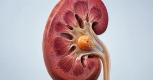
Left Flank Pain vs Back Pain: How to Tell Them Apart
Your left kidney and your lumbar muscles sit inches apart, but they produce pain that behaves nothing alike. Here are the six checks I use in clinic to separate left flank pain vs back pain before any scan is ordered.

Left flank pain vs back pain is a distinction most men try to make at 2 a.m., pressing on their own side and hoping the answer is a pulled muscle. Usually it is. But your left kidney sits only a few centimeters from the muscles you strained deadlifting, and the two produce pain that overlaps on a map while behaving nothing alike once you know what to test. The difference is not where the pain sits. It is what the pain does — whether it arrives in waves, whether changing position alters it, whether pressing one spot reproduces it, and whether anything has changed in your urine. In clinic I can separate the two with three questions and a urine dipstick before any scan is ordered. This article gives you the same six checks, the causes that are specific to the left side, and the situations where the correct response is an emergency room rather than a heat pack. For the wider picture on stone disease, start with the Kidney Stones Hub.
Key Takeaways
- Renal colic ignores position. If sitting, standing, or lying differently changes the pain, muscle or joint is the far more likely source.
- Pain travelling from the left flank down into the groin or testicle follows the ureter, not a nerve root — that pattern belongs to a stone.
- Blood is absent from the urine dipstick in roughly 10 to 30 percent of confirmed ureteric stones, so a clean sample does not clear you.
- A temperature of 38°C (100.4°F) or higher alongside left flank pain is a same-day emergency: an obstructed, infected kidney needs drainage within hours, not antibiotics alone.
- New left flank or back pain in a man over 60 who smokes or has smoked warrants an abdominal aortic aneurysm check before anything else is considered.
Where the Flank Ends and the Back Begins
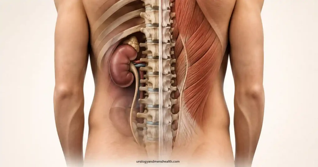
The flank is the strip of body wall between the bottom of your 12th rib and the top of your iliac crest, sitting to the side of the thick paraspinal muscles that run down either side of your spine. On most adult men it is about a hand’s width tall. Put your thumb on your lowest rib and your little finger on your hip bone, to the side of the spine, and you have covered it.
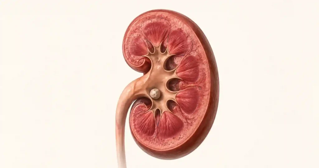
Your kidneys sit behind the abdominal cavity, packed in fat, tucked partly under the lower ribs. The left kidney sits higher than the right — roughly the level of the 11th rib to the second lumbar vertebra — because there is no liver above it pushing it down. That extra height matters: left kidney pain is often felt higher and further under the rib margin than men expect, which is exactly why they mistake it for a rib or muscle problem.
Directly behind the kidney sit the erector spinae and quadratus lumborum muscles, the thoracolumbar fascia, and the facet joints of the lumbar spine. These are the structures that strain when you lift badly, sit for nine hours, or sleep at an angle. Anatomically they are only two to five centimeters (about one to two inches) from the kidney’s back surface. The organs are neighbours. The pain they generate is not.
The reason they feel so similar is that kidney pain is visceral pain. It travels along sympathetic nerve fibers that enter the spinal cord around T10 to L1, and the brain has no precise map for those signals. So it reports something vague, deep, and broad — “somewhere in my left side” — rather than a point you can put a finger on. Muscular pain, by contrast, is somatic pain: sharply localized, reproducible, and tied to movement. That single mechanical difference generates almost every distinguishing sign below.
Left Flank Pain vs Back Pain: Six Differences That Actually Separate Them
These are the six checks I run through before touching a scan request. You can run them on yourself in about two minutes.
1. The shape of the pain over time
Renal colic comes in waves. It builds over 15 to 30 minutes to something close to unbearable, holds, then eases to a dull background ache before building again. That rhythm reflects the ureter contracting against an obstruction. Muscular pain is either constant and dull, or it spikes only when you move. A pain that has been identical in intensity for three days is rarely a stone.
2. What happens when you change position
This is the single most useful question. Lie flat. Roll onto the opposite side. Sit up and lean forward. If any of that reliably makes the pain better or worse, you are dealing with muscle, fascia, joint, or nerve root. Renal colic is indifferent to position. Men with a ureteric stone tell me they tried everything and nothing helped, which is precisely the diagnostic point. I go through this comparison in more depth in my breakdown of how stone pain differs from ordinary lumbar back pain.
3. How you behave during an episode
Men in renal colic pace, rock, and cannot stay still. Men with peritoneal irritation or a muscle tear stay very still, because movement hurts. Emergency physicians use this at the door: the restless patient is the one with the stone.
4. Where the pain travels
Radiation is close to a signature. Pain that runs from the left flank forward and down into the groin, the base of the penis, or the left testicle is following the ureter, and that is renal until proven otherwise. Pain that travels into the buttock and down the back of the leg below the knee is following a lumbar nerve root and is spinal. Pain that wraps around the trunk in a single band, at one level, is a thoracic nerve — and if it is burning and one-sided, watch for a shingles rash over the next few days.
5. What your body does alongside the pain
Nausea, vomiting, cold sweating, and pallor are autonomic responses to visceral pain. They come with stones, obstruction, and infection. A strained quadratus lumborum does not make you vomit or drench your shirt. If those symptoms are present, the source is an organ.
6. What is happening in your urine
Visible blood, pink or tea-colored urine, burning, a sudden need to pass urine every twenty minutes, or a drop in the volume you are producing all point at the urinary tract. A stone sitting at the junction of the ureter and the bladder classically produces urgency and burning that gets mistaken for an infection. Muscular back pain changes nothing about how you pass urine.
One more physical sign worth knowing: if firm finger pressure on a specific spot in the muscle beside your spine reproduces exactly the pain you have been feeling, that is your answer. Kidneys are cushioned by fat behind the peritoneum and are almost never tender to fingertip pressure. What a clinician tests instead is costovertebral angle tenderness — a flat thump over the angle between the 12th rib and the spine, which jars the whole kidney rather than compressing a muscle.
Kidney Causes of Left Flank Pain
A stone in the left ureter is the most common cause I see. Stones lodge at three narrow points: where the renal pelvis funnels into the ureter, where the ureter crosses the pelvic brim over the iliac vessels, and where it tunnels into the bladder wall. The last of these accounts for most of the emergency presentations, and it is the one that mimics a urine infection. Roughly two thirds of stones under 5 mm (about 1/5 inch) pass without intervention, while the odds fall substantially between 5 and 10 mm (about 2/5 inch) [1]. The full picture of how stones form, present, and get treated is set out in my complete guide to kidney stones.
Pyelonephritis — infection that has climbed into the kidney itself — produces flank pain that is constant rather than colicky, with fever, shaking chills, and marked costovertebral angle tenderness [4]. In men this is never a routine diagnosis. The male urethra is long enough that bacteria rarely reach the kidney without a structural reason: a stone, obstruction, incomplete bladder emptying, or an enlarged prostate. Any man diagnosed with an upper urinary tract infection needs an anatomical explanation, not just a prescription. I set out that workup in the guide to UTI in men and when to worry.
Obstruction without a stone is easy to miss. A narrowing at the ureteropelvic junction can stay silent for years and then produce a dull left flank ache an hour or two after a large fluid load — several beers, or a litre of water before a run. The kidney distends, the pain builds, and it settles once the fluid clears. Men often attribute this to alcohol rather than plumbing.
Bleeding into a kidney cyst gives sudden, sharp, one-sided flank pain, sometimes with visible blood in the urine, and usually settles over a few days. Kidney tumors, by contrast, are typically painless until late — which is why unexplained visible blood in the urine matters far more than pain as a warning sign. Renal infarction and renal vein thrombosis are rare but produce severe, abrupt, constant flank pain that does not follow a colicky rhythm, most often in men with atrial fibrillation, clotting disorders, or heavy protein loss in the urine.
Not sure how urgent this is? Run the Acute Stone Pain Triage Tool →Flank Pain That Might Be a Stone? Know What the Next 72 Hours Should Look Like
Enter your email below to receive Dr. Khalid’s complete Passing a Kidney Stone: What to Expect as a free, printable PDF.
Non-Kidney Causes on the Left Side
The left flank has a different neighbourhood from the right, and the list changes accordingly.
- Muscle and joint — quadratus lumborum or erector spinae strain, a facet joint irritation at L1 to L3, or a costovertebral joint sprain at the 11th or 12th rib. Reproducible on palpation, worse with extension or rotation, better with rest and heat. This is the majority of cases.
- Shingles before the rash appears — burning, intensely sensitive skin in a single band on one side, with pain preceding the blisters by two to four days. Men are frequently investigated for stones during that window.
- Diverticulitis — inflammation of pouches in the sigmoid colon, causing lower left-sided pain with fever and a change in bowel habit. It can refer backwards into the left flank.
- Splenic problems — infarction or an enlarged spleen produces left upper quadrant pain that can refer to the left shoulder tip. Uncommon, but the spleen has no equivalent on the right.
- Pancreatitis — pain in the upper abdomen boring straight through to the back, worse lying flat, better leaning forward. That forward-leaning relief is a strong clue.
- Lower lobe pneumonia or pleurisy — sharp pain worse on deep breath or cough, with the source above the diaphragm rather than below it.
- Abdominal aortic aneurysm — the diagnosis that must not be missed. New back or flank pain in a man over 60 with a smoking history, particularly with a pulsating sensation in the abdomen or any episode of feeling faint, requires urgent imaging [7].
In My Practice
A 34-year-old man came to clinic certain he had torn something in the gym. He had deadlifted heavy on the Saturday, woke on Sunday with left-sided pain, and by Tuesday was on his third heat pack. What made me pause was his answer to one question: nothing he did changed it. Not lying down, not stretching, not the heat. Pressing along his paraspinal muscles produced nothing. His dipstick showed microscopic blood, and the CT found a 6 mm stone at the left vesicoureteric junction. The deadlift was a coincidence he had built a whole diagnosis around.
When a man attributes flank pain to an activity but cannot make the pain change with position, the activity is almost always a coincidence rather than a cause.
Red Flags: When Left Flank Pain Needs the Emergency Room
Most left flank pain is not an emergency. These situations are, and the response to each is measured in hours rather than days.
Go to the Emergency Room Today
Any one of these alongside left flank pain means immediate assessment, not a next-available appointment:
- Fever of 38°C (100.4°F) or higher, or shaking chills. A blocked kidney that is also infected is a surgical emergency. It needs drainage with a stent or a nephrostomy tube, usually within hours. Antibiotics alone will not clear an obstructed system, and the deterioration can be rapid.
- You have only one working kidney, or a transplanted kidney, and the pain is on that side. There is no reserve.
- You have stopped producing urine, or the volume has fallen dramatically over 12 hours.
- Vomiting that stops you keeping fluids or painkillers down. Dehydration on top of obstruction accelerates kidney injury.
- Sudden severe pain with faintness, a pulsating abdominal mass, or a history of smoking and high blood pressure over the age of 60. This is an aneurysm assessment, and it takes priority over everything else on this page.
- Weakness in the legs, numbness across the saddle area, or a sudden inability to pass urine at all. These are spinal cord and cauda equina warning signs.
Two further situations need a doctor within the week rather than the hour: visible blood in the urine at any age with or without pain, and flank pain that has persisted beyond 72 hours without a clear mechanical explanation. Painless visible blood in the urine in particular should never be written off as a stone that has passed — it requires imaging of the upper tracts and a look inside the bladder.
How I Work Up Left Flank Pain in Clinic
The sequence is deliberately cheap before it becomes expensive.
History and the position test first. If the pain changes reliably with movement or posture and there are no urinary symptoms, the probability of a stone drops sharply and the workup shifts to a musculoskeletal one. The American College of Physicians is explicit that imaging for uncomplicated low back pain within the first six weeks does not improve outcomes and exposes patients to unnecessary radiation [5], and the same guidance body recommends starting with heat, movement, and non-drug measures rather than scans [6].
Urine dipstick and microscopy. Blood in the urine supports a stone but does not confirm one, and its absence does not exclude one — around 10 to 30 percent of men with a proven ureteric stone have no detectable blood on testing, most often when the ureter is completely blocked or the stone has been static for several days [3]. White cells and nitrites shift the concern towards infection.
Bloods. Creatinine and eGFR to check kidney function, plus white cell count and CRP if infection is on the table.
Imaging, chosen rather than reflexive. Non-contrast CT of the kidneys, ureters, and bladder detects stones with near-complete accuracy and remains the definitive test. But a large randomized trial published in the New England Journal of Medicine in 2014 found that starting with ultrasound produced no worse outcomes — no more complications, no more missed serious diagnoses — with substantially less radiation [2]. For a young man with a first episode, ultrasound first is a reasonable and often better starting point. For recurrent stones, an unclear picture, or anyone over 50 with new symptoms, I go straight to CT.
What to actually say at your appointment. Do not ask for “a scan.” Say this instead: “My pain does not change with position, it radiates into my groin, and I had nausea with it. Is a non-contrast CT KUB appropriate, or should we start with an ultrasound?” That sentence names the pattern, names the test, and moves the consultation forward in one step. If a stone is found, ask for its size in millimeters and its exact location, because those two numbers determine everything that happens next — the kidney stone passage calculator converts them into a realistic estimate of whether it will pass on its own and how long that is likely to take [8].
If the workup points away from the kidney, the plan for the first two weeks is specific: stay moving rather than resting in bed, apply heat for 15 to 20 minutes two or three times a day, use regular anti-inflammatories if you have no contraindication, and book physiotherapy if the pain has not improved by day 14. Track whether the pain is changing week to week. Pain that is static or worsening at four weeks, or that has developed any of the red flags above, gets re-assessed rather than re-treated.
Frequently Asked Questions
How do doctors tell left flank pain vs back pain apart in the emergency room?
We test three things: whether the pain changes with position, whether pressing on the muscle reproduces it, and what the urine dipstick shows. Renal colic ignores position, is not reproduced by finger pressure on the back, and usually shows blood in the urine. Muscular pain does the opposite on all three. If the answers point to the kidney, imaging follows, as covered in my breakdown of stone pain versus lumbar back pain.
Can left flank pain be a kidney stone if there is no blood in my urine?
Yes. Around 10 to 30 percent of men with a confirmed ureteric stone have no blood detectable on dipstick, usually because the stone is completely obstructing the ureter or has been static for several days. A negative dipstick lowers the probability but does not clear you. If the pain pattern fits colic, ask for imaging rather than accepting a normal urine test as the final answer. My complete kidney stone guide sets out the full workup.
Why does my left flank pain get worse when I twist or press on the area?
Reproducible tenderness is the strongest single sign that the source is muscle, fascia, or a facet joint rather than the kidney. The kidney sits behind the peritoneum, cushioned by fat, and fingertip pressure on the back rarely reaches it. Sharp pain that fires when you press one specific spot, twist, or arch backwards points to the thoracolumbar muscles. If you are still unsure, the acute stone pain triage tool walks through the same checks.
How long does left flank pain from a kidney stone usually last?
Each wave of colic typically builds and eases over 20 to 60 minutes, then repeats while the stone moves. Most stones under 5 mm (about 1/5 inch) pass within one to three weeks. Pain that stays constant for days without any wave pattern, or that persists after you believe the stone has passed, needs re-imaging rather than more painkillers. The kidney stone passage calculator gives a size-based estimate of the odds and timeline.
Can left flank pain come from an infection rather than a stone?
Yes. Infection that has reached the left kidney causes flank pain with fever, chills, and often nausea, and the tenderness is usually constant rather than colicky. In men an upper urinary tract infection is never routine and always needs a structural explanation, because the male urethra is long enough that bacteria rarely climb that far without an underlying cause. The article on UTI in men and when to worry explains which investigations should follow.
References
- American Urological Association / Endourological Society. Surgical Management of Stones: AUA/Endourology Society Guideline. AUA
- Smith-Bindman R, Aubin C, Bailitz J, et al. Ultrasonography versus computed tomography for suspected nephrolithiasis. N Engl J Med. 2014;371(12):1100-1110. PubMed
- Bove P, Kaplan D, Dalrymple N, et al. Reexamining the value of hematuria testing in patients with acute flank pain. J Urol. 1999;162(3 Pt 1):685-687. PubMed
- Gupta K, Hooton TM, Naber KG, et al. International clinical practice guidelines for the treatment of acute uncomplicated cystitis and pyelonephritis. Clin Infect Dis. 2011;52(5):e103-e120. PubMed
- Chou R, Qaseem A, Owens DK, Shekelle P. Diagnostic imaging for low back pain: advice for high-value health care from the American College of Physicians. Ann Intern Med. 2011;154(3):181-189. PubMed
- Qaseem A, Wilt TJ, McLean RM, Forciea MA. Noninvasive treatments for acute, subacute, and chronic low back pain: a clinical practice guideline from the American College of Physicians. Ann Intern Med. 2017;166(7):514-530. PubMed
- Chaikof EL, Dalman RL, Eskandari MK, et al. The Society for Vascular Surgery practice guidelines on the care of patients with an abdominal aortic aneurysm. J Vasc Surg. 2018;67(1):2-77. PubMed
- National Institute of Diabetes and Digestive and Kidney Diseases. Kidney Stones. NIDDK
Dr. Muhammad Khalid
MBBS · FCPS (Urology) · MCPS (General Surgery) · CHPE · CRSM · IMC #539472
Specialist urologist with 11+ years of clinical experience across tertiary teaching hospitals. Trained at Lady Reading Hospital and Khyber Teaching Hospital, Peshawar. Author or co-author of five peer-reviewed international publications, including in Cureus and the World Journal of Surgery. Procedural expertise: URS, PCNL, RIRS, TURP, TURBT, and major open urological surgery. Full profile →
This article is for educational purposes only and does not constitute medical advice. Always consult your physician or urologist for diagnosis and treatment decisions specific to your condition.