Bladder Cancer Symptoms, Stages & Diagnosis: A Guide
Painless blood in urine — especially in a man over 50 — is bladder cancer until a urologist proves otherwise. Here's how the symptoms, staging, and diagnosis actually work, and why the first missed clue costs the most.

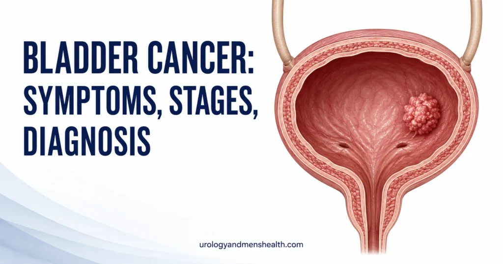
Most bladder cancer symptoms start with a single dismissed clue: a streak of blood in the urine that disappears within a day. I have seen too many men present in clinic six months later — when the painless hematuria came back, then came back again — only to find a muscle-invasive tumor that could have been a stage Ta paper-thin growth six months earlier. Painless blood in urine in a man over 50 is bladder cancer until a urologist proves otherwise. That single rule, applied early, is the single biggest determinant of how this disease turns out. This guide walks through the symptoms, the staging system, and the diagnostic pathway as I actually use them in clinic — and the moments where most delays happen. For wider context, see the full Surgery & Recovery Hub.
Key Takeaways
- Painless visible blood in urine (hematuria) is the presenting symptom in roughly 80% of bladder cancer cases and warrants urgent urology referral, regardless of how brief the episode was.
- Bladder cancer staging hinges on one cut-off: whether the tumor has invaded the bladder muscle (T2 and above) or stayed superficial (Ta, T1, CIS). That single line determines whether treatment is local or radical.
- Cystoscopy is the only reliable way to rule bladder cancer in or out. Urine tests and imaging support it; they do not replace it.
- Smoking is responsible for around half of all bladder cancers in men — the single largest modifiable risk factor on the list.
The Symptoms Most Men Ignore (And Why)
The hallmark of bladder cancer is painless, visible hematuria — urine that is pink, red, or tea-colored, with no burning, no flank pain, and no obvious trigger. About 8 in 10 patients with bladder cancer present this way [1]. The pattern that catches people out is its intermittency: blood appears for a day, sometimes a single void, then disappears for weeks. The tumor is still there. The bleeding stopped because a small vessel clotted off, not because the cause resolved.
The other symptoms are quieter and less specific. Microscopic hematuria — blood cells visible only on a urine dipstick or microscopy — is the second most common presentation, especially in patients picked up during routine screening or workup for another problem. Some patients describe urinary frequency, urgency, or a burning sensation that mimics a UTI but does not clear with antibiotics. This pattern matters: a recurrent UTI in a man, especially over 50, that does not culture an organism or that returns immediately after antibiotics finishes, should trigger urology referral, not another antibiotic course.
Less commonly, larger or locally advanced tumors cause flank pain (when a tumor at the trigone obstructs the ureter and backs urine up into the kidney), pelvic pain, or unintentional weight loss. By the time these symptoms appear, the cancer is usually no longer non-muscle invasive. For a wider breakdown of every cause of hematuria — not just cancer — see our guide on blood in urine and the 7 causes of hematuria in men.
In My Practice
A 62-year-old retired painter came to see me about a six-month history of “occasional pink urine” his primary care doctor had attributed to dehydration. Three urine dipsticks over that period had shown trace blood; no one had imaged him or sent him for cystoscopy. By the time I scoped him, he had a 3 cm papillary tumor on the right lateral wall — fortunately still Ta on TURBT histology. He was lucky. The painter’s history of solvent exposure was the second clue; the painless hematuria was the first.
Painless visible hematuria, even once, even in a young man, earns a urology referral — not a repeat dipstick in six weeks.
Who Actually Gets Bladder Cancer
Bladder cancer is the fourth most common cancer in American men, with the American Cancer Society estimating roughly 63,000 new male cases in 2024 [2]. Men are 3 to 4 times more likely to develop it than women, and the median age at diagnosis sits at 73. It is rare under 40, but rare is not the same as impossible — younger smokers and patients with occupational exposures do present, and a “you’re too young” response to visible hematuria has caused more than one delayed diagnosis in my own practice.
The risk factors with the strongest evidence are:
- Smoking. The single biggest preventable cause. Current smokers carry roughly 4 times the bladder cancer risk of never-smokers, and smoking accounts for around half of all male bladder cancers [3]. Risk falls after quitting but never returns to baseline.
- Occupational chemical exposure. Aromatic amines used in dye, rubber, leather, textile, paint, and aluminum industries are well-established carcinogens. Hairdressers, painters, truck drivers, and chemical plant workers carry measurably higher rates.
- Age over 55. Risk rises steeply from the late 50s onward.
- Male sex. Hormonal and exposure differences explain most of the gap.
- Chronic bladder inflammation. Long-term indwelling catheters, recurrent UTIs, and schistosomiasis (rare in the US, common in parts of Africa and the Middle East) all raise risk — schistosomiasis specifically for squamous cell bladder cancer.
- Prior pelvic radiation (for example, for prostate cancer 10-20 years earlier).
- Family history and certain genetic syndromes (Lynch syndrome), though these account for a small minority of cases.
Diet, fluid intake, and BMI have weaker evidence as risk factors. The headline action item from this list is unambiguous: if you smoke, the most powerful thing you can do for your bladder is stop. Risk reduction is measurable within 5 years.
Bladder Cancer Stages: What the Numbers Actually Mean
Bladder cancer staging uses the TNM system, but the clinically useful translation is simpler. Every bladder cancer falls into one of three boxes, and the box determines almost everything about treatment.
Box 1 — Non-muscle invasive bladder cancer (NMIBC)
This is the bladder wall sandwich’s outer two layers only. Roughly 75% of newly diagnosed bladder cancers sit here. It includes:
- Ta — papillary tumor confined to the urothelium (the bladder’s inner lining). Lowest risk.
- T1 — tumor has invaded the lamina propria (the connective tissue layer just under the lining), but not the muscle.
- CIS (carcinoma in situ) — flat, high-grade tumor confined to the urothelium. Looks like a velvety red patch on cystoscopy. Behaviorally aggressive despite being “non-invasive.”
NMIBC is treated by local resection (TURBT) and bladder-sparing therapy. Five-year survival for low-risk Ta tumors exceeds 90%, but recurrence is common — 50-70% within 5 years — which is why surveillance cystoscopy continues for years.
Box 2 — Muscle-invasive bladder cancer (MIBC)
The tumor has broken through into the bladder’s muscle layer (muscularis propria) or beyond. This is the pivot point at which treatment becomes radical.
- T2 — invades muscularis propria.
- T3 — invades perivesical fat (tissue around the bladder).
- T4a — invades adjacent organs (prostate, uterus, vagina).
Standard treatment is neoadjuvant cisplatin-based chemotherapy followed by radical cystectomy (removal of the bladder), or bladder-preserving trimodal therapy (maximal TURBT plus concurrent chemo-radiation) in selected patients. Five-year survival drops to roughly 50% at T2 and lower at T3-T4 [2].
Box 3 — Metastatic disease
The cancer has spread to regional lymph nodes (N1-N3), distant lymph nodes, or distant organs (M1) — most commonly lung, liver, and bone. T4b disease (invasion of pelvic or abdominal wall) is treated alongside metastatic disease. Treatment is systemic: platinum-based chemotherapy first-line, with immune checkpoint inhibitors (pembrolizumab, nivolumab) playing an increasing role in patients who progress on or cannot tolerate chemotherapy. Five-year survival is around 8-15% in this group.
The TNM letters also incorporate N (regional lymph node involvement, 0-3) and M (distant metastasis, 0 or 1). Grade — separate from stage — describes how abnormal the cells look under the microscope (low-grade vs high-grade). Two tumors at the same stage but different grades behave very differently; high-grade T1 cancer, in particular, behaves more like muscle-invasive disease and is often managed more aggressively than its stage alone would suggest.
How Bladder Cancer Is Actually Diagnosed
The diagnostic pathway is well-defined and unchanged across AUA, EAU, and NICE guidelines [4]. When a patient presents with hematuria, a urologist works through three steps, each of which answers a different question.
Step 1 — Urine tests (screening, not confirmatory)
Urinalysis confirms hematuria objectively (more than 3 red blood cells per high-power field). Urine culture rules out infection as a competing cause. Urine cytology looks for malignant cells shed into the urine — it is highly specific for high-grade disease and CIS but misses about 30-50% of low-grade tumors. A negative cytology does not rule cancer out. Urine-based molecular markers (UroVysion, NMP22, ImmunoCyt) exist but are not standalone replacements for cystoscopy in current US practice.
Step 2 — Upper tract imaging
Bladder cancer’s cousins — upper tract urothelial carcinoma of the renal pelvis and ureter — present with the same symptom and need ruling out. CT urogram (a CT scan with intravenous contrast that captures the urinary tract in three phases) is the standard of care. It also assesses the bladder wall, perivesical fat, regional lymph nodes, and the kidneys. In patients with contraindications to contrast, MR urogram is the alternative. The Urine Color Decoder tool can help readers interpret their own urine color while waiting for these results — see our urine color guide.
Step 3 — Cystoscopy
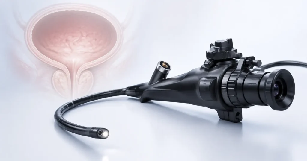
This is the diagnostic test. A thin flexible cystoscope is passed through the urethra into the bladder under local anesthetic, taking about 5 minutes in clinic. The urologist inspects the entire urothelial surface — the trigone, lateral walls, dome, and bladder neck — and photographs anything abnormal. If a tumor is seen (or suspected, as in red patches that may be CIS), the next step is examination under general anesthetic plus TURBT (transurethral resection of bladder tumor), which both removes the tumor and provides the histology that establishes stage and grade. The full procedure flow is covered in our deep dive on TURBT — the first procedure in bladder cancer treatment.
If TURBT histology returns muscle-invasive disease, the next step is staging imaging of the chest and pelvis (CT chest + abdomen + pelvis), plus a bone scan or PET-CT if symptoms suggest distant spread. From that, the multidisciplinary team — urologist, oncologist, radiologist, pathologist — chooses the treatment path.
Treatment Depends Entirely on the Stage
Treatment splits along the muscle-invasion line described above. This guide is about diagnosis rather than treatment, so the goal here is to give you a clear map of what is decided when, not a procedural walkthrough.
For NMIBC (Ta, T1, CIS): The first treatment is TURBT, which both removes the visible tumor and stages the disease. Based on the AUA/SUO risk stratification, the urologist then adds intravesical therapy: a single post-TURBT dose of mitomycin chemotherapy for low-risk Ta, or a 6-week induction course of BCG immunotherapy for high-risk Ta, T1, and CIS — BCG remains the single most effective intravesical agent for high-risk disease and reduces both recurrence and progression [5]. Surveillance cystoscopy follows for at least 5 years; for high-risk disease, it continues indefinitely.
For MIBC (T2-T4a, node-negative): Standard is neoadjuvant cisplatin-based chemotherapy (usually 3-4 cycles) followed by radical cystectomy with pelvic lymph node dissection. The bladder is removed; a urinary diversion is created from a segment of small or large bowel — either an ileal conduit (urine drains to a stoma bag) or an orthotopic neobladder (a continent reservoir reconnected to the urethra, allowing roughly normal voiding). In carefully selected patients — solitary tumor, complete TURBT possible, no widespread CIS, normal bladder function — trimodal therapy (maximal TURBT + chemo + radiation) preserves the bladder with comparable cancer-specific survival in good responders.
For metastatic disease: First-line is platinum-based combination chemotherapy (cisplatin + gemcitabine, or carboplatin + gemcitabine in patients ineligible for cisplatin). Maintenance immunotherapy with avelumab is increasingly used after a response to chemo. Second-line and beyond uses immune checkpoint inhibitors (pembrolizumab, nivolumab) and antibody-drug conjugates (enfortumab vedotin). The treatment landscape here is genuinely changing year on year, and a current oncology referral is essential. For a wider view of how urological cancer surgery is approached generally, our guide to urological surgical decision-making covers the principles that apply across stone and oncology procedures, and our Urology Surgery Comparison Tool lets you see how cystectomy compares to other major urological procedures.
When to See a Urologist (Don’t Wait)
The single most useful piece of advice I give in clinic is also the simplest: if you ever see blood in your urine, even once, even briefly, and especially if there is no pain, that finding needs a urologist’s eyes — not a wait-and-see appointment six weeks later. The cost of urgent referral is a clinic visit and possibly a cystoscopy. The cost of waiting is, in too many cases, an extra stage of disease.
Red Flags — See a Urologist Within 2 Weeks
Any one of the following warrants urgent referral, per AUA microscopic hematuria guidance and NICE NG12 cancer pathway criteria:
- Any episode of visible (gross) hematuria in an adult, regardless of how brief or whether pain was present.
- Microscopic hematuria in anyone over 35 with a smoking history, occupational chemical exposure, or other bladder cancer risk factors.
- Recurrent UTIs in a man that do not culture an organism or return immediately after a finished antibiotic course.
- New irritative urinary symptoms (frequency, urgency, suprapubic discomfort) without an obvious cause, especially over age 50.
- Flank pain combined with hematuria — concerns for either upper tract urothelial cancer or a trigonal bladder tumor obstructing the ureter.
Ask your primary care doctor to refer you for cystoscopy and CT urogram. If symptoms are severe or persistent, go to the emergency room — significant clot retention or obstruction is a urological emergency.
Bladder cancer caught at Ta or T1 is a different disease — clinically and emotionally — from bladder cancer caught at T2. The diagnostic pathway is short, mostly outpatient, and decisive. The cost of using it early is small. The cost of not using it is the thing this whole guide is built to help you avoid. For broader context on cancer screening across the male urological system, see our guide to prostate cancer screening — the screening principles are different, but the patient-level habits (knowing what to watch for, acting on the first clue) are the same.
Frequently Asked Questions
Can bladder cancer symptoms come and go before diagnosis?
Yes — and this is exactly why so many bladder cancers are diagnosed later than they need to be. Painless hematuria is classically intermittent: blood appears for a day or a single void, then the urine clears for weeks. The tumor is still there; the bleed simply stopped because a small surface vessel clotted off. A single episode of visible blood in urine — even if the next ten urine samples are clear — is enough to warrant urology referral and cystoscopy. See our detailed hematuria causes guide for the full differential.
How is bladder cancer staged once it’s diagnosed?
Staging uses the TNM system, but clinically it splits into three categories: non-muscle invasive (Ta, T1, CIS — the tumor sits in the bladder lining only), muscle invasive (T2 to T4a — the tumor has broken into the bladder muscle or surrounding fat), and metastatic disease. The split between non-muscle and muscle invasive determines whether treatment can be local (TURBT plus intravesical therapy) or has to be radical (cystectomy or trimodal chemo-radiation). Final staging requires TURBT histology plus, for muscle-invasive disease, CT chest, abdomen, and pelvis.
What are the early warning signs of bladder cancer in men?
The dominant early sign is painless visible hematuria — pink, red, or tea-colored urine without burning, flank pain, or fever. About 80% of bladder cancers present this way. Less common early signs include microscopic hematuria found on a routine urinalysis, recurrent urinary infections that do not culture an organism or that return immediately after antibiotics, and irritative symptoms (frequency, urgency, suprapubic discomfort) mimicking a UTI. Weight loss and flank or pelvic pain are late signs and usually mean the cancer is already locally advanced.
Is bladder cancer curable if caught early?
Yes — and the gap between early-stage and late-stage outcomes is one of the widest in solid-organ urology. Five-year survival for low-risk Ta non-muscle invasive bladder cancer treated with TURBT and intravesical therapy exceeds 90%. Five-year survival for muscle-invasive disease treated with neoadjuvant chemo and cystectomy is around 50%. Five-year survival for metastatic disease is roughly 8-15%. The pivot point is whether the cancer has reached the bladder muscle — which is exactly why early hematuria workup matters so much.
Does smoking really cause that much bladder cancer?
It is the single largest preventable cause. Current smokers carry roughly 4 times the risk of never-smokers, and smoking accounts for approximately half of all male bladder cancers in the US. The carcinogens — aromatic amines and polycyclic aromatic hydrocarbons — are absorbed from tobacco smoke, processed by the liver, and excreted in urine, where they sit in contact with the bladder lining. Quitting reduces risk measurably within 5 years, although risk does not return to never-smoker baseline. If you have other risk factors (occupational exposure, age over 55, family history), quitting is the single highest-impact action available.
How is cystoscopy actually performed — is it painful?
Flexible cystoscopy in clinic is uncomfortable, not painful. The cystoscope is a thin lubricated tube (about the diameter of a pencil) passed through the urethra into the bladder under topical lidocaine gel. Men feel a stretching sensation and a brief sting as the scope crosses the external sphincter; the procedure itself takes about 5 minutes. Most patients return to normal activity the same day, with a day or two of mild burning on urination afterward. If a tumor is found, the next step is TURBT under general anesthetic — covered in detail in our TURBT procedure guide.
References
- Lotan Y, Choueiri TK. Clinical presentation, diagnosis, and staging of bladder cancer. UpToDate. 2024. UpToDate
- American Cancer Society. Key Statistics for Bladder Cancer. cancer.org. 2024. ACS
- Freedman ND, Silverman DT, Hollenbeck AR, et al. Association between smoking and risk of bladder cancer among men and women. JAMA. 2011;306(7):737-745. PubMed
- Chang SS, Bochner BH, Chou R, et al. Treatment of Non-Metastatic Muscle-Invasive Bladder Cancer: AUA/ASCO/ASTRO/SUO Guideline. J Urol. 2017 (amended 2024). AUA
- Shelley MD, Mason MD, Kynaston H. Intravesical therapy for superficial bladder cancer: a systematic review of randomised trials and meta-analyses. Cochrane Database Syst Rev. 2010. Cochrane
Dr. Muhammad Khalid
MBBS · FCPS (Urology) · MCPS (General Surgery) · CHPE · CRSM · IMC #539472
Specialist urologist with 11+ years of clinical experience across tertiary teaching hospitals. Trained at Lady Reading Hospital and Khyber Teaching Hospital, Peshawar. Author or co-author of five peer-reviewed international publications, including in Cureus and the World Journal of Surgery. Procedural expertise: URS, PCNL, RIRS, TURP, TURBT, and major open urological surgery. Full profile →
This article is for educational purposes only and does not constitute medical advice. Always consult your physician or urologist for diagnosis and treatment decisions specific to your condition.