Hydronephrosis: What a Swollen Kidney on Your Scan Means
The word on your scan report tells me where urine is backing up, not why or how urgent it is. Here is how I sort a swollen kidney into watch, treat, or drain today.

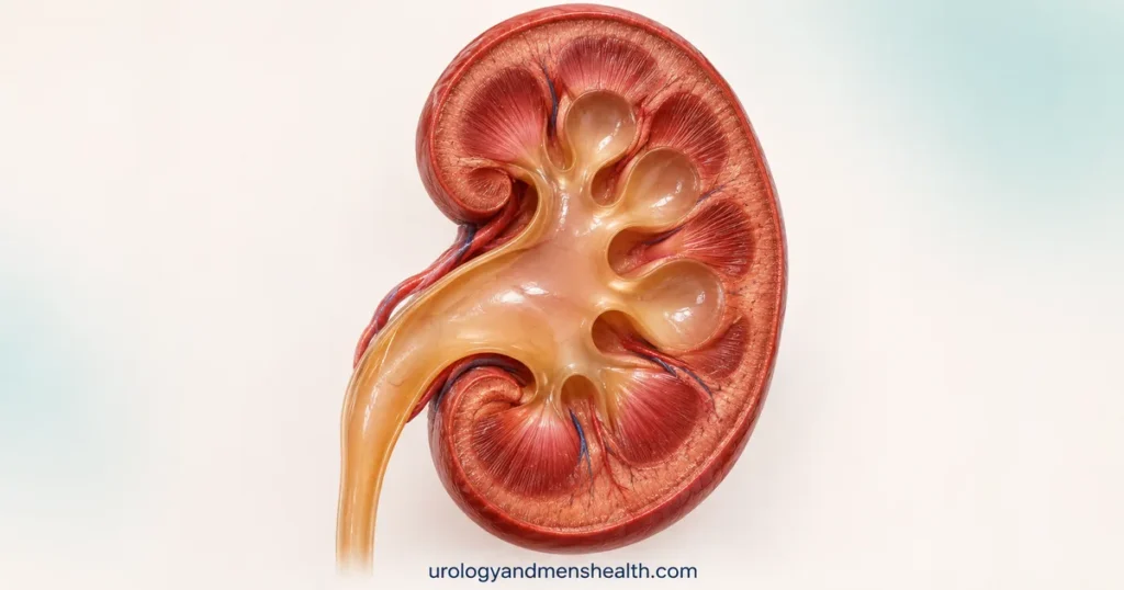
Hydronephrosis means your kidney is swollen with urine that cannot drain properly. If you are reading this because the word appeared on your ultrasound or CT report, the first thing to know is that it is a finding, not a diagnosis. It tells me where urine is backing up. It does not yet tell me why, how long it has been there, or whether the kidney is in danger. In my clinic, the same word on a report can mean a 4 mm stone that will pass by Friday, an enlarged prostate quietly holding back a liter of urine, or, less often, a tumor pressing on the ureter. The work is in sorting out which one you have and how urgent it is. This guide walks you through what the grades mean, what causes hydronephrosis in men, which tests come next, and when a swollen kidney needs drainage today rather than an appointment next month. For the wider picture of kidney blockages and stones, see the Kidney Stones Hub.
Key Takeaways
- Hydronephrosis with fever or chills is an emergency: an infected, blocked kidney needs drainage the same day, by stent or nephrostomy tube.
- A kidney stone is the most common cause of sudden one-sided hydronephrosis in men; an enlarged prostate is the most common cause of hydronephrosis in both kidneys.
- Mild hydronephrosis with normal kidney function and no pain is often watched with a repeat ultrasound rather than treated straight away.
- How much function the kidney recovers depends on how well it was working before the blockage was relieved, which is why delay matters.
What Hydronephrosis Means on Your Scan Report
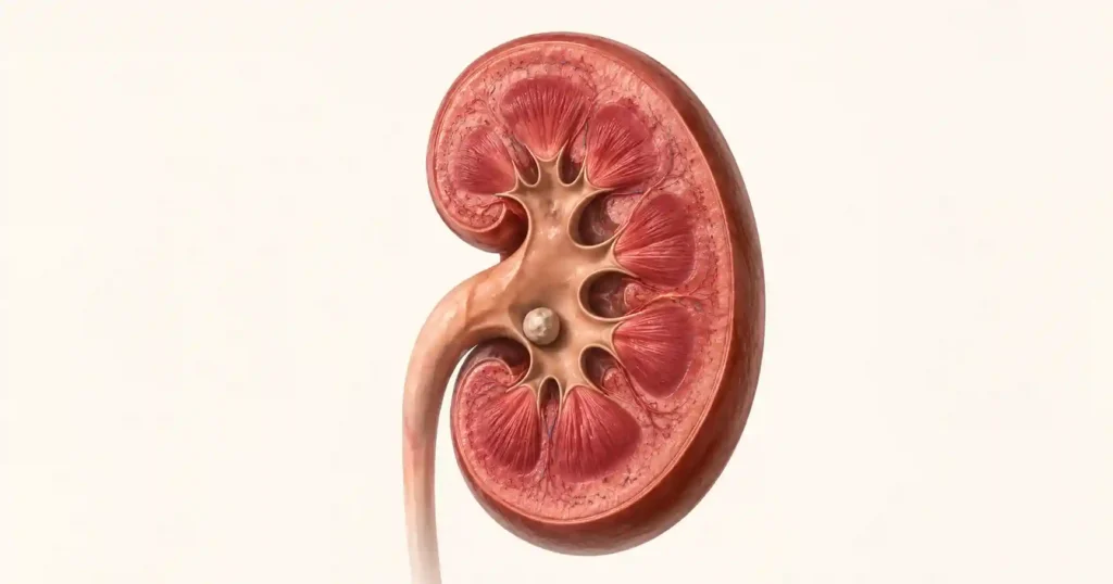
Your kidney makes urine continuously. It collects in a funnel-shaped space called the renal pelvis and its branches, the calyces, then drains down a narrow tube, the ureter, into the bladder. When anything slows that flow, pressure builds backward and the funnel balloons. That ballooning is hydronephrosis. If the ureter is also widened, the report may say hydroureteronephrosis, which tells me the blockage sits lower down, near or inside the bladder.
Think of a garden hose with a kink. The tap is still running, so the hose swells behind the kink. The kidney behaves the same way. In the first days, the swelling is mostly pressure. Over weeks, that pressure squeezes the working tissue around the funnel, called the cortex, and it starts to thin. A radiologist who writes cortical thinning is telling you the blockage has probably been there a while.
Two words on a report change how I read it. Unilateral means one kidney, which usually points to a problem in that ureter, such as a stone. Bilateral means both kidneys, which usually points to the bladder or prostate, because that is the one place both drainage routes share.
Grades of Hydronephrosis: Mild, Moderate, Severe
Most adult reports describe hydronephrosis as mild, moderate or severe. The grade describes the shape of the swelling, not how much damage has been done. A severely dilated kidney can still work well, and a mildly dilated one can be failing if the blockage is old. Grade has to be read alongside your blood tests and symptoms.
| Grade | What the scan shows | What I usually do next |
|---|---|---|
| Mild | Renal pelvis widened, calyces only slightly rounded | Find the cause; repeat ultrasound if no pain, fever or kidney function change |
| Moderate | Calyces clearly ballooned and blunted, cortex still normal thickness | CT or functional scan to locate the blockage and judge urgency |
| Severe | Large dilation with thinning of the kidney tissue | Drainage decision plus a renogram to measure how much function is left |
One trap catches people out. A full bladder can push back on the kidneys and make them look mildly dilated. If your scan was done with a bursting bladder, ask whether the kidneys were re-scanned after you emptied it. Mild dilation that disappears after voiding is usually not a true blockage.
What Causes Hydronephrosis in Men
I sort causes by where the blockage sits, because location predicts both urgency and treatment.
- Kidney stones. The most common cause of sudden, painful hydronephrosis on one side. A stone lodged in the ureter acts like the kink in the hose. Whether it passes on its own depends mostly on size and position; our kidney stone size chart shows pass rates by millimeter.
- Enlarged prostate and chronic urinary retention. When the prostate narrows the bladder outlet for years, the bladder stretches and stops emptying. Pressure then rises in both kidneys. This is the classic cause of bilateral hydronephrosis in men over 60, and it is often painless. The prostate side of this is covered in our guide to enlarged prostate treatment and surgery.
- Ureteropelvic junction (UPJ) obstruction. A narrowing where the kidney meets the ureter, usually present from birth. Some men only find out in their 30s or 40s, often after intermittent flank pain that gets worse after drinking a lot, such as after beer or coffee.
- Ureteric stricture. Scar tissue narrowing the ureter, sometimes after previous stone surgery, radiation or pelvic surgery.
- Tumors. A bladder cancer sitting over the ureter opening, prostate cancer, or enlarged lymph nodes in the abdomen can press on or block the ureter. This is why unexplained hydronephrosis in a smoker or a man with blood in his urine always gets imaging of the whole urinary tract.
- Retroperitoneal fibrosis. A rare condition where fibrous tissue wraps around the ureters, typically causing hydronephrosis on both sides.
In My Practice
A 68-year-old man was referred to me urgently with a report reading bilateral hydronephrosis and a creatinine of 265 umol/L (3.0 mg/dL). He was frightened about dialysis. He told me he passed urine fine, just often and in small amounts. A bladder scan showed well over a liter left behind after he had just been to the toilet. The small, frequent voids were overflow from a bladder that never emptied. A catheter went in that afternoon, and over the next ten days his creatinine fell steadily toward normal.
Bilateral hydronephrosis in an older man is a bladder question first: measure what is left after voiding before ordering anything more complex.
Hydronephrosis Symptoms: When It Hurts and When It Is Silent
Symptoms depend on how fast the blockage happened, not how big the kidney looks. A sudden blockage stretches the kidney capsule quickly, and stretch is what hurts. A slow blockage gives the kidney time to adapt, so it can swell enormously with no pain at all.
- Colicky flank pain spreading to the groin: the ureter is contracting hard against a stone. This is the acute picture.
- A dull ache in the side or back: steady pressure in a dilated kidney, more typical of UPJ narrowing or a slow-growing blockage.
- Nausea and vomiting: the kidney and gut share nerve pathways, so kidney stretch triggers the vomiting center.
- Weak stream, dribbling, getting up repeatedly at night: points to the bladder outlet and the prostate as the source of bilateral hydronephrosis.
- No symptoms: common when hydronephrosis is found by chance on a scan done for something else. Silent does not mean harmless; it can mean the kidney has been slowly losing function.
Pain from the kidney is easy to confuse with muscle pain in the back. Location, timing and what makes it worse help tell them apart; our page on flank pain versus back pain walks through the differences.
Go to the ER Today If
Hydronephrosis becomes an emergency when infection or kidney failure joins the blockage. Do not wait for a clinic appointment if you have:
- Fever of 38 C (100.4 F) or higher, shivering or chills, with flank pain or a known stone
- Hydronephrosis in your only working kidney, or in both kidneys at once
- Very little or no urine output over several hours
- Vomiting so persistent you cannot keep fluids or pain medication down
An infected, blocked kidney can turn into sepsis within hours. Antibiotics alone do not reliably clear infection trapped behind a blockage, which is why both the American Urological Association and the European Association of Urology treat urgent drainage as the priority in this situation [1][2].
Hydronephrosis on your scan? Get the stone action plan: what to ask, and when to go to the ER
Enter your email below to receive Dr. Khalid’s complete Passing a Kidney Stone: What to Expect as a free, printable PDF.
Tests After a Hydronephrosis Finding
The tests answer three questions in order: is it an emergency, where is the blockage, and how much is the kidney still doing.
- Creatinine and eGFR. A blood test showing how well both kidneys filter together. One blocked kidney can be hidden by a healthy partner, so a normal result does not rule out damage to one side. You can see where your number sits with our eGFR and CKD stage calculator, and our guide to high creatinine explains when a rise matters.
- Urinalysis and urine culture. To look for infection, which changes the urgency completely.
- Post-void bladder scan. A quick ultrasound after you urinate. If a large volume stays behind, the bladder is the problem.
- Non-contrast CT of the kidneys, ureters and bladder (CT KUB). The most accurate scan for finding a stone and its exact size. For an uncomplicated suspected stone, starting with ultrasound is reasonable: a large randomized trial of 2,759 emergency patients found that initial ultrasound cut radiation exposure without increasing missed serious diagnoses [3].
- CT urogram. CT with contrast, used when a tumor or stricture is possible, or when there is blood in the urine.
- Diuretic renogram (MAG3 scan). A nuclear medicine scan that measures how much each kidney contributes and whether urine truly cannot get out. This is the test that decides whether a chronically dilated kidney is worth repairing.
What to ask for: if your report says hydronephrosis and nobody has checked your creatinine and urine within the week, ask your primary care doctor for both, plus a post-void bladder scan if you are over 50. Bring the full report to your urologist, including the kidney size and any mention of cortical thinning.
Hydronephrosis Treatment: Stent, Nephrostomy or Watchful Waiting
Treatment has two parts: relieve the pressure, then fix the cause. Sometimes both happen at once; often they are separated by weeks.
Watchful waiting is right for a man with mild hydronephrosis, a small stone likely to pass, no fever and normal kidney function. Medication that relaxes the ureter may help the stone along; our guide to tamsulosin for passing kidney stones explains who benefits. I set a time limit: if the stone has not passed or the pain keeps returning, we intervene rather than letting a blocked kidney sit for weeks.
A ureteric stent is a thin flexible tube placed through the bladder up into the kidney, usually under a short anesthetic. It holds the ureter open so urine bypasses the blockage. Stents can be uncomfortable, and our article on managing stent pain covers what helps.
A percutaneous nephrostomy is a tube placed through the skin of the back directly into the kidney, usually by a radiologist under local anesthetic, draining into a bag. It is the preferred option when a man is too unwell for anesthesia or when a stent cannot pass the blockage. In a randomized trial in patients with an infected kidney blocked by a stone, stent and nephrostomy relieved the obstruction and infection equally well; neither led to faster recovery [4].
A urinary catheter is the first step when the bladder is the cause. After a long blockage is relieved, some men pass very large volumes of urine for a day or two, called post-obstructive diuresis, so fluid balance and blood tests are checked closely in that window. Definitive treatment then targets the cause: stone removal, prostate surgery, reconstruction of a UPJ narrowing (pyeloplasty), or cancer treatment.
Can a Kidney Recover From Hydronephrosis?
Usually yes, partly or fully, if the blockage is relieved in time. The best predictor is how well the kidney was working when the blockage was relieved. In a prospective study of 91 adults with one blocked kidney, a kidney that still filtered at least 10 mL/min on a renogram before treatment stabilized or improved afterward, while kidneys below that level did not recover [5]. Work from the Sindh Institute of Urology and Transplantation in Karachi found that how much urine drains through a nephrostomy tube in the first days predicts later recovery more reliably than imaging alone [6].
In practice this means two things. First, a stone causing complete blockage should not be left for weeks in the hope it passes; the longer the kidney is under pressure, the less comes back. Second, a kidney that contributes very little on a renogram, with a healthy kidney on the other side, may be better removed than repaired. That is a conversation for your urologist with the renogram result in hand. Expect a repeat ultrasound four to six weeks after the cause is treated to confirm the swelling has settled.
If you have a stone and want a personal estimate of whether it will pass on its own, try our kidney stone passage calculator.
Frequently Asked Questions About Hydronephrosis
Is mild hydronephrosis serious?
Mild hydronephrosis with no pain, no fever and normal creatinine is usually not urgent, but it still needs a cause. Ask for a repeat ultrasound after emptying your bladder, since a full bladder can mimic mild dilation. If it persists, a CT or renogram shows whether there is a real blockage. Pass rates for small stones are in our stone size chart.
Can hydronephrosis go away on its own?
Yes, when the cause clears. Hydronephrosis from a small stone usually resolves within days to weeks after the stone passes. Hydronephrosis from a fixed narrowing, chronic urinary retention or a tumor does not resolve until that cause is treated. A follow-up ultrasound four to six weeks later confirms the kidney has drained. Our Kidney Stones Hub covers stone-related causes in detail.
Why do both of my kidneys show hydronephrosis?
When both kidneys are swollen, the blockage is usually at or below the bladder, because that is the only point both kidneys share. In men over 60 the commonest cause is an enlarged prostate stopping the bladder from emptying. A post-void bladder scan and creatinine test come first. Our guide to enlarged prostate treatment explains what follows.
Do I need a stent or a nephrostomy for hydronephrosis with infection?
Either works. A randomized trial found stents and nephrostomy tubes relieved obstruction and infection equally well, so the choice depends on how unwell you are, whether you can have anesthesia, and what is causing the blockage. What matters is that an infected, blocked kidney is drained the same day. If you already have a stent, see our guide to stent pain.
Will my kidney function come back after hydronephrosis is treated?
Recovery depends mainly on how well the kidney was working before the blockage was relieved. Kidneys that still filter reasonably on a renogram usually stabilize or improve, while those with very little remaining function often do not. Ask for your creatinine to be repeated after treatment and track your eGFR with our CKD stage calculator.
References
- Assimos D, Krambeck A, Miller NL, et al. Surgical Management of Stones: American Urological Association/Endourological Society Guideline. American Urological Association. 2016. AUA
- European Association of Urology. EAU Guidelines on Urolithiasis. EAU Guidelines Office. EAU
- Smith-Bindman R, Aubin C, Bailitz J, et al. Ultrasonography versus computed tomography for suspected nephrolithiasis. N Engl J Med. 2014;371(12):1100-10. PubMed
- Pearle MS, Pierce HL, Miller GL, et al. Optimal method of urgent decompression of the collecting system for obstruction and infection due to ureteral calculi. J Urol. 1998;160(4):1260-4. PubMed
- Khalaf IM, Shokeir AA, El-Gyoushi FI, et al. Recoverability of renal function after treatment of adult patients with unilateral obstructive uropathy and normal contralateral kidney: a prospective study. Urology. 2004;64(4):664-8. PubMed
- Hussain M, Ali B, Ahmed S, et al. Prediction of renal function recovery in obstructive renal failure due to stones. J Pak Med Assoc. 1997;47(6):159-61. PubMed
Dr. Muhammad Khalid
MBBS · FCPS (Urology) · MCPS (General Surgery) · CHPE · CRSM · IMC #539472
Specialist urologist with 11+ years of clinical experience across tertiary teaching hospitals. Trained at Lady Reading Hospital and Khyber Teaching Hospital, Peshawar. Author or co-author of five peer-reviewed international publications, including in Cureus and the World Journal of Surgery. Procedural expertise: URS, PCNL, RIRS, TURP, TURBT, and major open urological surgery. Full profile →
This article is for educational purposes only and does not constitute medical advice. Always consult your physician or urologist for diagnosis and treatment decisions specific to your condition.