Imaging for Recurrent UTI in Men: Which Scans You Need
Most men referred for imaging for recurrent UTI get an ultrasound, and for most of them that is the right first scan. The harder question is which men need more — and what a normal result does not rule out.

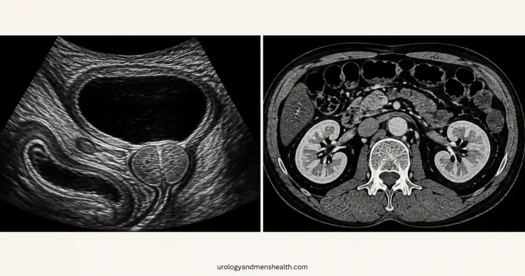
Imaging for recurrent UTI in men answers one question: is something in the urinary tract making these infections keep happening? In women, repeat infections are usually a bacterial and behavioral problem, and scanning rarely adds anything. In men it is the reverse. The male urethra is long, and prostatic secretions are mildly antibacterial, so bacteria that reach the bladder again and again usually have help — a bladder that does not empty, a stone acting as a reservoir, a narrowed urethra, or a prostate holding organisms between courses of antibiotics. That is why almost every man with two or more culture-proven infections in six months is scanned, and why which scan he is offered matters. Most men need one test. Some need three. A smaller group is told everything looks normal and keeps getting infections anyway — which is a finding in itself, not a dead end. This article covers which scan comes first, the specific triggers that justify moving to a CT urogram or cystoscopy, and what each result changes about treatment. For the wider picture, see our UTIs and Infections Hub.
Key Takeaways
- Two culture-proven infections in six months, or three in twelve, is the threshold at which a man’s urinary tract gets imaged — the working assumption is that something structural or functional is driving them.
- Renal and bladder ultrasound with a post-void residual is the correct first scan for nearly every man. It uses no radiation and finds the commonest cause, which is a bladder that does not empty.
- A CT urogram is triggered by specific findings — stone history, alkaline urine, fever that has not settled after 72 hours of antibiotics, blood in the urine that persists after clearance, or air in the urine — not by frustration with an ultrasound.
- Normal scans do not close the case. Chronic bacterial prostatitis, urethral stricture and small bladder tumors are diagnosed by culture, flow studies and cystoscopy, not by ultrasound.
Why Imaging for Recurrent UTI in Men Is Standard, Not Optional
Recurrent means two culture-proven infections within six months or three within twelve months. That definition matters, because it separates a man who has had one infection from a man whose urinary tract is repeatedly losing the same fight.
The contrast with women is instructive. The American College of Radiology’s 2026 update on recurrent lower urinary tract infection concluded that imaging is usually not appropriate for a woman with uncomplicated recurrent infections and no underlying risk factors [3]. Men sit at the other end of that scale. In a prospective cohort of men referred to urology with proven infection, 53 of 100 fully evaluated patients had an important urological abnormality, and the single commonest one — present in 34 of them — was a bladder that emptied poorly [4]. Roughly half of men investigated for infection have something wrong that a scan can show.
There is one honest exception. In a prospective series of 29 otherwise healthy men aged 16 to 45 admitted with a first episode of infection, ultrasound, urography and cystoscopy were normal in every single one [5]. A young man with one infection and a normal flow does not need a scan. A young man with three does. Age lowers the threshold rather than setting it — the trigger is recurrence, not birthday.
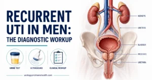
What imaging is looking for is a mechanical explanation, and there are only a handful: urine left behind after voiding, a stone that bacteria colonize, a pouch in the bladder wall that does not drain, a narrowing that slows flow, or a prostate that reseeds the bladder after each antibiotic course. The full diagnostic sequence — cultures, timing, flow studies, when to refer — is covered in our diagnostic workup for recurrent UTIs in men. This article stays on the scans.
In My Practice
A man in his late sixties came to me having had four infections in a year, each treated by his primary care doctor with a different antibiotic, each clearing and coming back within eight to ten weeks. Nobody had scanned him. His bladder ultrasound showed a post-void residual of 380 mL — he was carrying most of a bladderful of stagnant urine around all day, every day. His infections were not an antibiotic problem at all.
In men, a repeating infection is usually the symptom; the plumbing is the disease, and no antibiotic course fixes plumbing.
The First Scan: Renal and Bladder Ultrasound With a Post-Void Residual
Ask for it by its full name: renal and bladder ultrasound with post-void residual. The post-void residual is the part that gets left off request forms, and it is the part most likely to explain your infections. Without it, you have paid for two-thirds of a test.
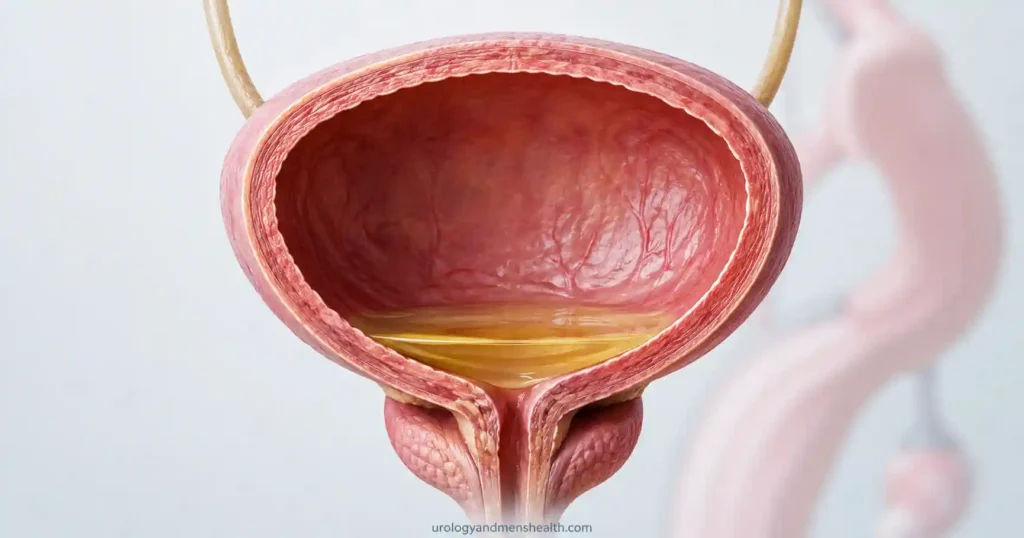
A single sitting gives your urologist five things: whether either kidney is swollen from obstruction (hydronephrosis), whether there are stones in the kidneys or bladder, whether the bladder wall has thickened and roughened from years of pushing against resistance, how large the prostate is, and how much urine is still sitting in the bladder after you have finished voiding.
That last number is the highest-yield measurement in the whole workup. Commonly used clinical thresholds put a residual under 50 mL (about 1.7 fl oz) in the normal range, 50–100 mL as borderline, and anything repeatedly above 100–150 mL (roughly 3.5–5 fl oz) as a genuine explanation for recurrence. Stagnant urine is a culture medium at body temperature. Bacteria that would have been flushed out by a complete void instead get several hours to multiply.
Two practical points on timing. Have the residual measured when you are free of symptoms, ideally at least two weeks after finishing antibiotics — an inflamed bladder voids abnormally and produces a residual that is not representative. And void normally on a normal urge, not on command with a barely full bladder, or the number is meaningless in the other direction. If you already have a residual figure and want to see where it sits, run it through our post-void residual danger estimator.
Ultrasound has real blind spots, and knowing them is what stops you from over-trusting a normal report. It misses small stones sitting in the ureter, it is poor at showing tumors of the kidney’s drainage system, it cannot see a urethral stricture at all, and it will not show a connection between bowel and bladder.
Recurrent UTIs in men: the step-by-step eradication protocol, including which scan to request first
Enter your email below to receive Dr. Khalid’s complete Recurrent UTI Eradication Protocol as a free, printable PDF.
When Ultrasound Is Not Enough: Triggers for CT, Cystoscopy and Flow Studies
A CT urogram is not a better ultrasound. It is a different test with a different purpose, and it carries a radiation dose in the range of several years of natural background exposure plus an iodinated contrast load that matters if your kidney function is reduced. European Association of Urology guidance is explicit that an extensive routine workup has a low diagnostic yield and should be reserved for atypical cases — but performed without delay when stone disease, obstruction or urothelial cancer is suspected [2]. The skill is in recognizing which case is atypical.
These are the triggers that move a man past ultrasound:
- Fever and flank pain that have not settled after 72 hours of an antibiotic the organism is sensitive to. That combination raises the possibility of a kidney abscess or an obstructed, infected kidney, and CT is the test that finds both.
- A history of kidney stones, a urine pH of 7.0 or above, or an eGFR of 40 or below. A validated clinical rule built from adults with febrile infection used exactly these three factors and ruled out urgent urological disorders with a negative predictive value of 99% when all three were absent — enough to cut imaging by around 40% without losing significant findings [6].
- Repeated growth of Proteus or another urease-producing organism alongside alkaline urine. Those bacteria manufacture the chemistry that grows struvite (infection) stones, which can fill a collecting system while an ultrasound reports little more than shadowing.
- Blood in the urine that persists once the infection has cleared. This crosses into a different pathway entirely — see the next section.
- Bubbles of air in the urine stream, fecal matter in the urine, or repeated growth of several organisms at once. That triad suggests a connection between bowel and bladder, usually from diverticular disease. Ultrasound will not show it; CT with rectal contrast will.
- A slow, straining stream, or previous catheterization, instrumentation or urethral infection. This points at a urethral stricture, which is diagnosed by flow rate plus cystoscopy or a retrograde urethrogram — never by ultrasound.
| Test | What it finds | What it misses |
|---|---|---|
| Renal and bladder ultrasound with post-void residual | Hydronephrosis, kidney and bladder stones, thickened or trabeculated bladder wall, diverticula, prostate size, incomplete emptying | Small ureteric stones, upper tract tumors, urethral strictures, bowel-to-bladder fistulas |
| CT urogram | Stones of any size, upper tract tumors, kidney abscess, fistulas, the exact level of an obstruction | Fine detail of the bladder lining; carries radiation and a contrast load |
| Flexible cystoscopy | Bladder tumors and stones, trabeculation, diverticular necks, urethral strictures, an obstructing prostate seen directly | Everything above the bladder — kidneys and ureters are invisible from inside |
| Uroflowmetry | An obstructed or underactive voiding pattern and a low peak flow rate | The cause of the pattern, which needs ultrasound or cystoscopy to identify |
| Transrectal prostate ultrasound | Prostatic abscess and prostatic cysts, in selected cases only | Chronic bacterial prostatitis itself, which is a culture diagnosis and not an imaging one |
| Retrograde urethrogram | The exact length and position of a urethral stricture before repair | Kidneys, bladder and prostate entirely |
One pathway deserves separating out. If blood is still present in your urine after the infection has been treated and cleared, you are no longer being investigated for infection — you are being investigated for a bladder or kidney tumor, and the rules change. The AUA and SUFU microhematuria guideline, amended in 2025, stratifies men by age and smoking history: under 40 with a minimal smoking history is low or negligible risk, 40 to 59 is intermediate, and 60 and over is high risk. High-risk patients should have cystoscopy plus axial upper tract imaging — a CT urogram — rather than an ultrasound [1].
What the Scan Result Actually Changes
A scan is only worth having if the result alters what happens next. Here is what each of the common findings changes.
High residual with an enlarged prostate
Treatment moves off antibiotics and onto the obstruction — an alpha blocker, a 5-alpha reductase inhibitor, or a procedure, depending on prostate size and how much bladder function is left. Ask your urologist directly: “if we fix the emptying, do you expect the infections to stop?” In men whose residual is the whole story, the honest answer is usually yes.
A bladder stone or an infection stone in the kidney
The stone comes out. This is not negotiable and it is not something antibiotics can work around — bacteria live inside the stone’s matrix where drug levels never reach. With struvite stones in particular, every fragment must be cleared, because residual fragments reseed the infection and the stone regrows. A stone found on imaging converts a medical problem into a surgical one.
A bladder diverticulum or a stricture
A diverticulum is a pouch of bladder wall that fills when you void and never fully drains — a permanent reservoir. A stricture is a narrowed segment that raises voiding pressure and leaves urine behind. Both are structural and both are repaired surgically when the infection burden justifies it. Neither responds to prophylactic antibiotics for longer than the antibiotics are taken.
Everything normal, and the same organism every time
This is the pattern that frustrates men most, and it has a name. When identical organisms relapse after each course, the prostate is acting as a sanctuary — most antibiotics penetrate prostatic tissue poorly, so a seven-day course clears the bladder and leaves the reservoir intact. Chronic bacterial prostatitis is diagnosed on culture rather than on any scan, and it is treated with four to six weeks of an antibiotic that actually gets into prostate tissue, most often a fluoroquinolone. EAU guidance reserves transrectal prostate ultrasound for selected cases where a prostatic abscess is suspected, not as a routine part of the workup [2]. If your scans are clean and your cultures keep growing the same bug, ask specifically whether you have had a prostate-directed course of adequate length — not whether you need another scan.
When to Go to the Emergency Room
Recurrent infection is an outpatient problem until it is not. Go to the emergency room the same day, rather than waiting for a scan appointment, if you develop any of the following:
- Fever above 38°C (100.4°F) with shaking chills alongside flank or back pain — an infected, obstructed kidney needs drainage within hours, not antibiotics within days
- Complete inability to pass urine despite the urge
- Confusion or unusual drowsiness in an older man with a urinary infection — this is often the only sign of sepsis
- Feeling faint on standing, a racing pulse, or a marked drop in urine output
Frequently Asked Questions
How many infections does a man need before a scan is justified?
Two culture-proven infections in six months, or three in twelve, is the working threshold. A single episode in a man under 40 with a normal stream rarely justifies imaging — a prospective series of 29 healthy young men admitted with a first infection found normal ultrasound, urography and cystoscopy in every one of them. Recurrence, not the first episode, is what triggers the scan.
Is an ultrasound enough, or should I push for a CT urogram?
For most men, ultrasound with a post-void residual is the right and sufficient first test. Imaging for recurrent UTI in men escalates on specific triggers: a stone history, urine pH of 7.0 or above, reduced kidney function, fever that has not settled after 72 hours of appropriate antibiotics, blood in the urine that persists after clearance, or air in the urine. Without one of those, a CT adds radiation and contrast without adding answers.
My post-void residual came back at 180 mL — is that what is causing my infections?
Very likely, yes. A residual of 180 mL is well above the 50 mL considered normal and above the 100 to 150 mL range at which incomplete emptying becomes a credible explanation for recurrence. Have it repeated once while you are infection-free to confirm it is your baseline rather than a one-off, then ask what is causing it. Our guide to what a post-void residual result means covers the thresholds in detail.
Why does my urologist want a cystoscopy when both my scans were normal?
Because ultrasound and CT both look at the bladder from outside. A cystoscope looks at the lining from inside, and that is the only way to see a urethral stricture, a small flat bladder tumor, the narrow neck of a diverticulum, or a stone hidden behind an enlarged prostatic lobe. In men with repeated infections and clean cross-sectional imaging, cystoscopy is where the diagnosis is most often made.
I still had blood in my urine after the infection cleared — does that change which scan I need?
It changes the whole pathway. Blood that persists once the infection is treated is investigated as possible urothelial cancer rather than as infection. Under the 2025 AUA and SUFU microhematuria guideline, men aged 60 and over fall into the high-risk group and should have cystoscopy plus axial upper tract imaging. Our article on blood in the urine in men over 60 explains why age shifts the workup.
References
- Barocas DA, Lotan Y, Matulewicz RS, et al. Updates to Microhematuria: AUA/SUFU Guideline (2025). J Urol. 2025;213(5):547-557. AUA
- EAU Guidelines on Urological Infections. European Association of Urology; 2026. EAU
- Expert Panel on Urological Imaging. ACR Appropriateness Criteria: Recurrent Lower Urinary Tract Infections in Females: Update 2026. J Am Coll Radiol. 2026. JACR
- Andrews SJ, Brooks PT, Hanbury DC, et al. Ultrasonography and abdominal radiography versus intravenous urography in investigation of urinary tract infection in men: prospective incident cohort study. BMJ. 2002;324(7335):454-456. PubMed
- Abarbanel J, Engelstein D, Lask D, Livne PM. Urinary tract infection in men younger than 45 years of age: is there a need for urologic investigation? Urology. 2003;62(1):27-29. PubMed
- van Nieuwkoop C, Hoppe BPC, Bonten TN, et al. Predicting the need for radiologic imaging in adults with febrile urinary tract infection. Clin Infect Dis. 2010;51(11):1266-1272. PubMed
Dr. Muhammad Khalid
MBBS · FCPS (Urology) · MCPS (General Surgery) · CHPE · CRSM · IMC #539472
Specialist urologist with 11+ years of clinical experience across tertiary teaching hospitals. Trained at Lady Reading Hospital and Khyber Teaching Hospital, Peshawar. Author or co-author of five peer-reviewed international publications, including in Cureus and the World Journal of Surgery. Procedural expertise: URS, PCNL, RIRS, TURP, TURBT, and major open urological surgery. Full profile →
This article is for educational purposes only and does not constitute medical advice. Always consult your physician or urologist for diagnosis and treatment decisions specific to your condition.