Shockwave Lithotripsy Candidacy: Are You a Good Fit?
Shockwave lithotripsy candidacy is decided by four numbers already sitting on your CT scan: stone size, location, density in Hounsfield units, and skin-to-stone distance. Here is how I read them before offering ESWL.

Shockwave lithotripsy candidacy is decided long before you lie down on the machine — it is decided by four numbers that are already sitting on your CT scan. ESWL (extracorporeal shockwave lithotripsy) is the only stone treatment that never puts an instrument inside your body, and that makes it the most attractive option on paper. It is also the option most frequently offered to the wrong patient. In clinic I regularly see the consequence: men who have had two or three sessions elsewhere, still have the stone, and were never told that their scan predicted failure from the start. The procedure is not the problem. Patient selection is. This article walks through exactly what I check before I offer ESWL — stone size, stone location, stone density in Hounsfield units, and skin-to-stone distance — plus the situations where shockwaves are simply off the table. For the wider picture of stone disease, start with our Kidney Stones Hub.
Key Takeaways
- ESWL does not remove your stone. It breaks it into fragments you still have to pass yourself, which is why the pain often arrives in the days after the procedure rather than during it.
- Stones under 10 mm (about 2/5 inch) sitting in the upper or middle calyx or the proximal ureter are the sweet spot. In the Lower Pole I trial, only 21% of patients with lower pole stones above 10 mm were stone-free after ESWL.
- Density decides more than most patients realize: in a Toronto series, ESWL succeeded 91% of the time when the stone measured under 900 HU with a skin-to-stone distance under 9 cm — and only 41% when both numbers were unfavorable.
- Skin-to-stone distance predicts failure independently of BMI. A distance above 10 cm (about 4 inches) on CT is a warning sign, not a formality.
- Pregnancy, an untreated urinary infection, uncorrected clotting problems, and an obstruction below the stone are all reasons ESWL should not go ahead.
How Shockwave Lithotripsy Actually Breaks a Stone
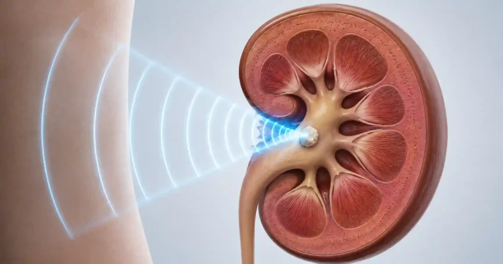
A lithotripter generates a pressure wave outside your body — electromagnetically, electrohydraulically, or piezoelectrically depending on the machine — and focuses it through a water or gel cushion onto a single point inside the kidney. Your urologist lines that focal point up with the stone using either X-ray fluoroscopy or ultrasound. Nothing enters the body.
Two separate forces then do the work. The first is direct mechanical stress: the wave compresses the front face of the stone and, as it exits the far side, creates tension that propagates cracks backward through the crystal structure. Think of a hammer blow travelling through a brick and splitting it from the inside out. The second is cavitation — microscopic bubbles form in the surrounding fluid, then collapse violently against the stone surface, firing tiny jets that pit and erode it.
A typical session delivers 2,000 to 3,000 shocks over 45 to 60 minutes, usually under sedation or light general anesthesia, and you go home the same day. Delivery rate matters: shocks given at 60 to 90 per minute fragment stones more effectively than faster rates, because slower delivery allows cavitation bubbles to clear between waves.
Here is the part patients consistently misunderstand. The machine’s job ends when the stone is in pieces. Your ureter does the rest. ESWL converts one large stone into a shower of small ones that then have to travel down a tube roughly 3 mm wide. That is why a man can leave the lithotripsy suite feeling fine and be in significant pain four days later.
The Four Numbers That Decide Shockwave Lithotripsy Candidacy
Every one of these is measurable on the non-contrast CT scan you have almost certainly already had. Ask for them by name.
1. Stone size
The 2026 AUA Surgical Management of Kidney and Ureteral Stones guideline treats both ESWL and ureteroscopy as reasonable first-line choices for renal stones up to 20 mm (about 4/5 inch) [1]. But performance falls off steeply as size climbs. Under 10 mm, ESWL earns its place. Between 10 and 20 mm, the honest conversation is about how many sessions you are willing to accept. Above 20 mm, percutaneous nephrolithotomy is the answer, and the 2026 update added mini-PCNL as an option for stones of 1 to 3 cm — for the 1 to 2 cm band it may be offered ahead of ureteroscopy on stone-free rate grounds.
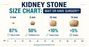
If you want to see where your specific stone sits against the wait-versus-treat threshold, the kidney stone size chart and treatment guide lays out the millimeter bands in detail.
2. Stone location
Upper and middle calyces drain downhill into the renal pelvis, so fragments clear well. Proximal ureteral stones also respond, because the stone is fixed in a narrow channel and the shock wave has a stable target. Distal ureteral stones are a different case — ureteroscopy reaches them directly and produces higher single-session clearance. And the lower pole is the weak point, which gets its own section below.
3. Stone density in Hounsfield units
Hounsfield units (HU) measure how much X-ray energy the stone absorbs — effectively, how hard it is. In a St. Michael’s Hospital series of 111 patients, stones under 900 HU fragmented reliably while denser stones did not, and this held independently of stone size, location and BMI [4]. A 2023 systematic review reached the same conclusion across the wider literature [5]. My working threshold in clinic: under 900 HU I offer ESWL confidently, 900 to 1,000 HU I discuss the odds openly, and above 1,000 HU I usually steer toward ureteroscopy.
4. Skin-to-stone distance
This is the straight-line distance from the skin surface to the stone, averaged across three measurements on axial CT. Every centimeter of tissue the shock wave crosses costs it energy. Pareek and colleagues showed that skin-to-stone distance predicts stone-free status after ESWL independently of BMI, with distances at or above 10 cm marking the failure zone for lower pole stones [3]. Two men with the same BMI can differ by 3 cm depending on where their fat is distributed — which is exactly why BMI alone is a poor screening tool here.
The two numbers compound. When Perks stratified patients into four groups, the ESWL success rate ran 91%, 79%, 58% and 41% across the combinations of density and distance — from under 900 HU with distance under 9 cm at the top, to 900 HU or more with distance of 9 cm or more at the bottom [4]. A man in the bottom group is being offered a coin flip.
Work through the same four variables I use in clinic with the Kidney Stone Surgery Selector →Why Lower Pole Stones Are ESWL’s Weak Spot
The lower pole calyx sits at the bottom of the kidney. When you are upright, it is the dependent compartment — the basement. Fragments created there do not drain downhill into the renal pelvis; they have to travel upward through a narrow channel called the infundibulum before they can leave the kidney at all.
Three anatomical measurements determine whether that journey happens:
- Infundibulopelvic angle — the angle between the infundibulum and the renal pelvis. A sharp, acute angle traps fragments.
- Infundibular length — longer than 3 cm (about 1.2 inches) and fragments stall partway.
- Infundibular width — narrower than 5 mm and even small fragments jam.
The Lower Pole I trial randomized 128 patients with lower pole stones to ESWL or percutaneous removal. At three months, the stone-free rate was 37% for ESWL versus 95% for percutaneous nephrolithotomy. For stones larger than 10 mm the ESWL figure collapsed to 21%, and there were nine treatment failures in the ESWL arm against none in the percutaneous arm [2]. That trial is over two decades old and lithotripter technology has moved on, but the anatomical problem it identified has not — gravity has not been updated.
This does not make lower pole ESWL wrong in every case. A 6 mm lower pole stone at 700 HU with a wide, short infundibulum in a slim patient is still a sensible ESWL target, and recovery is easier than any alternative. Above 10 mm in the lower pole, though, I move to flexible ureteroscopy or mini-PCNL. If you are weighing those against each other, the comparison of URS, RIRS and PCNL for stone disease sets out what each procedure demands of you.
Passing Stone Fragments After Lithotripsy: What to Expect Week by Week
Enter your email below to receive Dr. Khalid’s complete Passing a Kidney Stone: What to Expect as a free, printable PDF.
Stone Composition: Which Stones Simply Do Not Break
Hardness is not just a number on a scan — it reflects what the stone is made of. Some crystal structures shatter; others absorb the energy and stay intact.
Resistant to shockwaves: calcium oxalate monohydrate (whewellite), brushite (calcium hydrogen phosphate), and cystine. These have dense, uniform crystal lattices with few internal flaws for a crack to travel along. Cystine stones in particular can survive multiple full sessions unchanged.
Fragments readily: calcium oxalate dihydrate (weddellite), struvite, and uric acid. These are more porous and internally irregular, so cracks propagate easily.
Uric acid stones deserve a caveat. They fragment well, but they are radiolucent — invisible on plain X-ray — so targeting needs ultrasound or contrast. More to the point, most uric acid stones will dissolve with urinary alkalinization using potassium citrate. If your stone is uric acid, the right first move is often a prescription rather than a procedure. Density is a useful clue here: uric acid stones typically read in the 300 to 500 HU range, while cystine and brushite sit well above 1,000 HU. The stone composition identifier helps you interpret a previous stone analysis report, and the full breakdown of kidney stone types explains how each one forms.
If you have ever caught and submitted a stone for analysis, bring that report to your consultation. It is the cheapest predictive test in the entire workup and it is routinely left in a drawer at home.
In My Practice
A man in his forties came to me after two ESWL sessions at another center for a 9 mm lower pole stone. He was frustrated and assumed the machine had failed him. I pulled up his original CT: mean attenuation 1,180 HU, skin-to-stone distance 12.4 cm, and the stone sitting at the base of a long, narrow lower pole infundibulum. Every one of those numbers had been on the scan before the first session. His stone came out with a single flexible ureteroscopy three weeks later.
When all four predictive numbers point the wrong way, a second ESWL session is not persistence — it is a repeat of a decision that was already wrong.
When ESWL Is Off the Table: Contraindications
Some ESWL contraindications are absolute. No amount of favorable stone anatomy overrides them:
- Pregnancy — shock waves and the fluoroscopy used to aim them are both unacceptable risks to a fetus.
- Untreated urinary infection — fragmenting an infected stone releases bacteria and endotoxin into an obstructed system. Urine culture must be sterile or treated first.
- Uncorrected bleeding disorder or active anticoagulation — the kidney absorbs shock wave energy and bleeds. Note the asymmetry in the 2026 AUA guidance: low-dose aspirin may now be continued through PCNL in selected patients, but anticoagulation still has to be held before ESWL [1].
- Obstruction below the stone — a stricture or another stone downstream means fragments have nowhere to go. You will have converted one problem into a hundred.
- Aortic or renal artery aneurysm in the shock path — a straightforward mechanical exclusion.
Relative contraindications require a judgment call rather than a refusal: severe obesity where the stone cannot be brought into the machine’s focal zone, severe skeletal deformity preventing positioning, uncontrolled hypertension (which raises hematoma risk), stones inside a calyceal diverticulum with no drainage route, and implanted cardiac devices, which usually proceed safely after clearance from the device team.
One practical change worth knowing: the 2026 AUA guideline states that clinicians may omit prophylactic antibiotics before shockwave lithotripsy in patients without infection [1], and the 2026 EAU urolithiasis guideline revised its own antibiotic prophylaxis section along similar lines [6]. If you are not offered antibiotics, that is current practice, not an oversight — provided your urine culture was clear.
When to Go to the ER After ESWL
Fragment passage causes cramping pain that settles with oral analgesia. These do not:
- Fever above 38°C (100.4°F) or shaking chills — an infected, obstructed system after lithotripsy can become septic within hours
- Pain that does not respond to your prescribed medication, or that comes with persistent vomiting
- Passing no urine at all, or a sudden drop in output — particularly if you have one working kidney
- Heavy visible blood with clots, or bleeding that continues beyond 48 hours
- Steady, unrelenting flank pain a week or more after treatment, which can mean a column of fragments has stacked up in the ureter
What to Ask Before You Agree to Shockwave Lithotripsy
Take these six questions into the consultation. Every one has an answer already recorded on your scan or in your chart.
- What is my stone’s maximum diameter in millimeters, and exactly which calyx or ureteral segment is it in? If the answer is lower pole and above 10 mm, ask directly why ureteroscopy is not being offered first.
- What is the mean attenuation in Hounsfield units? Above 1,000 HU, ask what the expected single-session success rate is for that density at this center.
- What is my skin-to-stone distance? If it has not been measured, ask for it to be measured before the date is booked.
- Do I have a previous stone analysis on file? Cystine or brushite on a past report should redirect the plan entirely.
- Will I be stented? Routine pre-stenting is not required for ESWL, but a stent changes your recovery substantially and you should know in advance.
- What is the plan if this session does not work, and when will you image me to find out? Expect a specific interval — typically two to four weeks — and a specific next step.
On that last point, the 2026 AUA guideline supports a repeat ESWL session after an incomplete response, then a transition to ureteroscopy if the retreatment also fails [1]. My own rule is firmer: two sessions with imaging between them, and never a third on optimism alone. Each session carries anesthetic exposure, renal parenchymal trauma and weeks of your life.
And once the stone is clear, the work is not finished. Lithotripsy treats the stone; it does nothing about the metabolic reason you built one. Ask for a 24-hour urine collection six to eight weeks after clearance and send any fragment you catch for analysis — the metabolic workup for recurrent kidney stones explains what those tests look for and what changes in response to the results.
Frequently Asked Questions
What actually decides shockwave lithotripsy candidacy — my stone or my body?
Both, and they are weighed together. Stone size, location and density come from your CT scan; skin-to-stone distance is a body measurement taken from the same scan. A small, low-density stone can still fail in a heavier patient because the shock wave loses energy crossing tissue before it arrives. Our kidney stone surgery selector works through the same variables I use in clinic.
Why did my urologist rule out ESWL for a lower pole stone?
Because gravity works against you there. The lower pole calyx sits at the bottom of the kidney, so fragments must travel upward through a narrow infundibulum to reach the renal pelvis and drain. In the Lower Pole I trial, only 21 percent of patients with lower pole stones above 10 mm were stone-free after ESWL. Flexible ureteroscopy or PCNL clears these far more reliably.
What Hounsfield unit reading is too high for shockwave lithotripsy?
There is no single cut-off, but the evidence clusters between 900 and 1,000 HU. Below 900 HU stones fragment well; above 1,000 HU I usually recommend ureteroscopy instead. Composition explains why — cystine, brushite and calcium oxalate monohydrate are dense and resist shockwaves. If you have a previous stone analysis, our stone composition identifier helps you read it.
Does being overweight rule out shockwave lithotripsy?
Not automatically. What matters is skin-to-stone distance measured on CT, not BMI on its own. Two men with identical BMIs can differ by 3 cm depending on where their fat sits. Above roughly 10 cm the failure rate climbs sharply because the wave dissipates before reaching the stone. Stone size still drives the decision too, as our stone size chart explains.
How many ESWL sessions should I accept before switching to another procedure?
The 2026 AUA guideline supports a repeat session after an incomplete response, then a move to ureteroscopy if that retreatment also fails. My own rule is two sessions with imaging in between, never a third on hope alone. If the stone has not fragmented after two, the decision framework comparing ESWL, ureteroscopy and PCNL is where to go next.
My stone cleared after ESWL — why did another one form two years later?
Because lithotripsy treats the stone, not the reason you formed it. Recurrence within five to ten years is common when the underlying cause is never identified. After clearance I ask for a 24-hour urine collection plus a stone analysis, then target the specific abnormality found. The metabolic workup for recurrent stones explains what those tests look for.
References
- Pearle MS, Matlaga BR, Antonelli JA, et al. Surgical Management of Kidney and Ureteral Stones: AUA Guideline (2026). Part II: Evaluation and Treatment of Patients With Kidney and/or Ureteral Stones. J Urol. 2026;215(2):124-131. PubMed
- Albala DM, Assimos DG, Clayman RV, et al. Lower pole I: a prospective randomized trial of extracorporeal shock wave lithotripsy and percutaneous nephrostolithotomy for lower pole nephrolithiasis — initial results. J Urol. 2001;166(6):2072-2080. PubMed
- Pareek G, Hedican SP, Lee FT Jr, Nakada SY. Shock wave lithotripsy success determined by skin-to-stone distance on computed tomography. Urology. 2005;66(5):941-944. PubMed
- Perks AE, Schuler TD, Lee J, et al. Stone attenuation and skin-to-stone distance on computed tomography predicts for stone fragmentation by shock wave lithotripsy. Urology. 2008;72(4):765-769. PubMed
- Garg M, Johnson H, Lee SM, Rai BP, Somani B, Philip J. Role of Hounsfield Unit in Predicting Outcomes of Shock Wave Lithotripsy for Renal Calculi: Outcomes of a Systematic Review. Curr Urol Rep. 2023;24(4):173-185. PubMed
- Skolarikos A, Jung H, Neisius A, et al. EAU Guidelines on Urolithiasis. Edn. presented at the EAU Annual Congress London 2026. European Association of Urology Guidelines Office, Arnhem. EAU
Dr. Muhammad Khalid
MBBS · FCPS (Urology) · MCPS (General Surgery) · CHPE · CRSM · IMC #539472
Specialist urologist with 11+ years of clinical experience across tertiary teaching hospitals. Trained at Lady Reading Hospital and Khyber Teaching Hospital, Peshawar. Author or co-author of five peer-reviewed international publications, including in Cureus and the World Journal of Surgery. Procedural expertise: URS, PCNL, RIRS, TURP, TURBT, and major open urological surgery. Full profile →
This article is for educational purposes only and does not constitute medical advice. Always consult your physician or urologist for diagnosis and treatment decisions specific to your condition.