Renal Artery Stenosis: Diagnosis and When a Stent Helps
Most men told they have renal artery stenosis assume a stent is the fix. Two large randomized trials say otherwise for the majority. Here's how I decide who genuinely benefits from opening the artery.

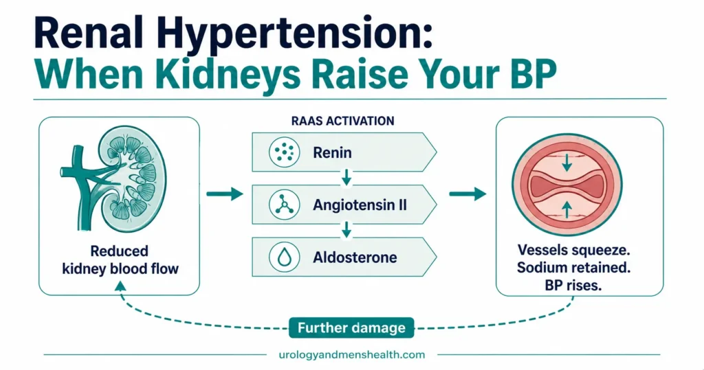
Renal artery stenosis is a narrowing of one or both of the arteries that carry blood to your kidneys, and it sits behind a small but meaningful share of blood pressure that refuses to come down. The kidney does not know why its blood supply has dropped. It responds the way it would respond to blood loss — by releasing renin, the hormone that switches on the renin-angiotensin system — and pressure climbs across the entire circulation. The narrowing is the problem, but the pressure is what gets measured, so the treatment keeps chasing the wrong target. Most men who hear this diagnosis assume the fix is mechanical: open the artery, place a stent, done. Two large randomized trials have shown that assumption is wrong for the majority of people with plaque-related disease. Below is what actually narrows the artery, which clinical clues should trigger testing, how the diagnosis is confirmed, and the specific situations where opening the artery still changes outcomes. For the wider picture on pressure and kidney function, see our Blood Pressure & Kidney Hub.
Key Takeaways
- About 90% of renal artery stenosis is atherosclerotic plaque sitting at the point where the renal artery leaves the aorta; most of the remainder is fibromuscular dysplasia, which behaves and responds differently.
- The clues that matter are specific: pressure still high on three or more drugs including a diuretic, a sharp creatinine rise within weeks of starting an ACE inhibitor or ARB, one kidney more than 1.5 cm (about 0.6 inch) smaller than the other, or sudden fluid on the lungs with a normally pumping heart.
- Duplex ultrasound is the first test. A peak systolic velocity above roughly 180–200 cm/s together with a renal-to-aortic ratio of 3.5 or higher points to a narrowing of 60% or more.
- In both the CORAL and ASTRAL trials, adding a stent to good medical therapy did not reduce death, heart attack, stroke, or progression to dialysis in atherosclerotic disease.
- Fibromuscular dysplasia is the opposite story: balloon angioplasty without a stent is first-line, and roughly a third of patients come off blood pressure medication entirely.
What Narrows the Renal Artery — and Why It Raises Your Blood Pressure
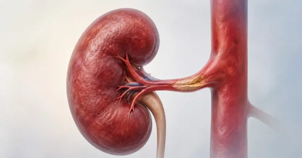
Each kidney is fed by a single main artery branching straight off the aorta. That artery is short, wide, and carries about a fifth of everything your heart pumps. It is also a favourite site for plaque, because the turbulence where a vessel leaves the aorta damages the lining over decades.
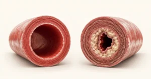
Roughly 90% of cases are atherosclerotic — the same cholesterol-laden plaque that narrows coronary and carotid arteries, deposited at the ostium where the renal artery originates [1]. If you have plaque here, you almost certainly have it elsewhere. That is the single most useful thing to understand about this diagnosis: it is a marker of body-wide vascular disease, not an isolated plumbing fault.
Most of the rest is fibromuscular dysplasia (FMD) — an abnormal overgrowth of cells in the artery wall that has nothing to do with cholesterol. It typically strikes women between 25 and 50, sits in the middle or far end of the artery rather than the origin, and produces a distinctive alternating narrow-wide pattern on angiography that radiologists call a string of beads [1].
The renin mechanism, in plain terms
The kidney has no way to distinguish “my artery is narrowed” from “the body is losing blood.” Both feel identical from inside the organ: less pressure arriving. So it does what evolution built it to do — it releases renin, which triggers a cascade that constricts blood vessels everywhere and tells the body to retain salt and water. The narrowed kidney gets the perfusion it wanted. Every other organ pays for it with elevated pressure. This is renovascular hypertension, and it is why the pressure is so stubborn: the kidney is actively defending it. For the broader mechanisms behind kidney-driven pressure, including the non-obstructive causes, see how renal hypertension develops and what changes the trajectory.
Whether one artery is narrowed or both matters enormously, and it changes what medication is safe. With one narrowed artery, the healthy kidney can still handle salt and water, and the picture is dominated by high pressure. With both narrowed — or with a single functioning kidney whose artery is narrowed — there is no compensating organ. Fluid accumulates, kidney function becomes fragile, and drugs that relax the blood vessel on the far side of the filter can tip function downward abruptly.
Renal Artery Stenosis Symptoms: The Clues That Should Trigger Testing
Here is the uncomfortable part: renal artery stenosis symptoms barely exist. The artery narrows silently over years. There is no flank pain, no change in urination, no ache. What you get instead is a set of laboratory and clinical patterns that a doctor has to actively recognise, because nothing about how you feel will point at the artery [1].
These are the patterns worth acting on:
- Pressure that stays above target on three or more drugs, one of which is a diuretic, taken as prescribed. This is the classic trigger.
- Hypertension that appears before age 30 or after age 55. Early onset suggests fibromuscular dysplasia; late onset suggests plaque.
- A creatinine rise of more than about 30% within two to four weeks of starting an ACE inhibitor or an ARB. This is close to a diagnostic signature for narrowing in both arteries, or in the artery to a solitary kidney.
- A size difference between the kidneys of more than 1.5 cm (about 0.6 inch) on any ultrasound or CT scan. The smaller kidney has been under-perfused for a long time.
- Kidney function that is declining without an obvious cause — no diabetes, no protein in the urine, no history of obstruction.
- A bruit — a whooshing sound heard with a stethoscope over the upper abdomen. Its presence is suggestive; its absence rules out nothing [1].
When to Go to the ER
One presentation of renal artery narrowing is a genuine emergency and is often mistaken for a heart attack. Go to the emergency room immediately if you develop:
- Sudden breathlessness with frothy sputum, coming on over minutes to hours, especially if you cannot lie flat. Fluid can flood the lungs abruptly in bilateral disease even when the heart is pumping normally.
- A sudden severe rise in blood pressure with headache, blurred vision, chest pain, or confusion.
- A sharp drop in urine output over 24 to 48 hours, particularly after starting a new blood pressure medication.
Tell the emergency team you have known or suspected renal artery stenosis. It changes which drugs they reach for.
Blood Pressure and Kidney Protection: the tests to request when your pressure will not come down
Enter your email below to receive Dr. Khalid’s complete Blood Pressure & Kidney Protection Guide as a free, printable PDF.
How Renal Artery Stenosis Is Diagnosed
Duplex ultrasound is the first test in almost every case. It combines standard imaging with Doppler measurement of how fast blood is moving, and it costs little, uses no contrast, and carries no radiation. A peak systolic velocity above roughly 180 to 200 cm/s in the renal artery, combined with a renal-to-aortic ratio of 3.5 or higher, indicates a narrowing of 60% or more [2]. The ratio matters because pressure alone can raise velocities throughout the abdomen — comparing the renal artery to the neighbouring aorta separates a true narrowing from generally fast flow.
The weakness of duplex is that it depends heavily on the person holding the probe. Body habitus, bowel gas, and accessory renal arteries all defeat it. A negative scan in a patient with strong clinical suspicion should not close the question.
CT angiography gives the clearest anatomy and is fast, but delivers a substantial contrast load — a genuine problem when the kidney you are investigating is already impaired. MR angiography avoids radiation and provides some functional information about flow, but gadolinium contrast is generally avoided when eGFR falls below 30. Both tests have a known tendency to make a narrowing look worse than it is [1].
Catheter angiography remains the reference standard, and in the same session a pressure wire can measure the actual gradient across the lesion and treatment can be delivered. That is precisely why it should not be a screening test. Once a catheter is in the renal artery and a narrowing is visible, the decision to treat it becomes very hard to resist — regardless of whether treating it will help. In practice, catheter angiography belongs at the point where you have already decided that intervention is on the table.
Check your kidney damage risk from your own BP readings, kidney history, and current medications →What to request: if your pressure remains above target on three drugs including a diuretic, ask your primary care doctor or a nephrologist specifically for a renal artery duplex ultrasound, and ask for renal function and electrolytes to be drawn on the same day so the numbers can be read together. If you have recently started an ACE inhibitor or ARB, ask for a creatinine and potassium check within one to two weeks of starting — not at the routine three-month review.
Medical Treatment Comes First — and for Most People, That Is the Whole Treatment
This is where patient expectation and evidence diverge most sharply. The intuition — a narrowed pipe should be reopened — is strong, and it was standard practice for years. Then the trials arrived.
The CORAL trial randomized 947 patients with atherosclerotic renal artery narrowing plus either systolic hypertension on two or more drugs or chronic kidney disease, assigning them to medical therapy alone or medical therapy plus a stent. There was no difference in the composite outcome of cardiovascular or renal death, heart attack, stroke, hospitalization for heart failure, progressive kidney failure, or need for dialysis. Systolic pressure was lower in the stent group by 2.3 mmHg — statistically detectable, clinically close to meaningless [3].
The ASTRAL trial had already reached the same conclusion with 806 patients, finding substantial procedural risk without matching benefit [4]. Extended follow-up of the ASTRAL cohort out to a median of 56 months found no difference in kidney outcomes, cardiovascular events, or survival by intention-to-treat [5]. Two large trials, long follow-up, consistent answer.
What “medical therapy” actually means here
The trials did not compare stenting against doing nothing. They compared it against a specific and demanding regimen, and understanding that regimen is what makes the result usable rather than discouraging. In CORAL it consisted of an angiotensin receptor blocker, a thiazide-type diuretic, amlodipine, atorvastatin, antiplatelet therapy, and guideline-level diabetes control [3]. Add smoking cessation, which does more for a renal artery than any procedure will.
ACE inhibitors and ARBs are the counter-intuitive part. They block the exact hormone system the narrowed kidney is using to drive your pressure, which makes them the logical drug — and they also reduce filtration pressure inside the kidney, which is why function can fall when both arteries are narrowed. The resolution is not avoidance but monitoring: start the drug, recheck kidney function within one to two weeks, and act on the number. The long-term damage that untreated pressure inflicts on the kidney is covered in detail in our guide to how high blood pressure damages kidneys before symptoms appear.
In My Practice
A man in his mid-sixties was referred to me after his creatinine jumped from 105 to 168 µmol/L (1.19 to 1.90 mg/dL) three weeks after his primary care doctor added ramipril for pressure that had not budged on two drugs. Duplex showed narrowing in both renal arteries. The referral question, written plainly at the bottom of the letter, was “does he need a stent?” He did not. Stopping the ramipril brought his creatinine back to baseline within a month. Amlodipine and a thiazide, with atorvastatin and low-dose aspirin added, held him at 134/78 mmHg for the next two years, and his kidney function has not moved since.
A creatinine rise after starting an ACE inhibitor is one of the most reliable clues that both arteries are narrowed — but it is a diagnostic finding, not an indication to open the artery.
Who Actually Benefits From a Renal Artery Stent
“Stenting does not help on average” is not the same as “stenting never helps.” The trials enrolled patients with moderate narrowing and reasonably stable disease. They deliberately excluded, or barely included, the small group in whom the artery is doing obvious active harm. That group still exists, and a renal artery stent is still the right answer for them.
- Flash pulmonary edema with preserved heart function. Sudden fluid on the lungs in someone with narrowing in both arteries, when the heart itself is pumping normally, is the strongest single indication to revascularize.
- Kidney function falling fast and demonstrably tied to the artery — particularly when the narrowing sits in the artery to a solitary functioning kidney.
- Genuinely refractory hypertension: still above target on four or more drugs at proper doses, with confirmed adherence, and a lesion severe enough to produce a real pressure gradient across it.
- Intolerance of the drugs that would otherwise control the situation, leaving no viable medical route.
Notice what unites these. In each, the narrowing is producing an acute or accelerating problem, not a chronically elevated number. Severity of the narrowing on a scan is not on the list. A 70% lesion in a man whose pressure is controlled on three drugs and whose creatinine has been flat for four years is a scan finding, not a treatment target.
Fibromuscular dysplasia is a different disease with a different answer
Everything above applies to plaque-related disease. FMD inverts it. Here, balloon angioplasty without a stent is first-line treatment, and the aim is not merely to lower pressure but to cure it. Across pooled data, roughly 36% of patients achieve blood pressure below 140/90 mmHg with no medication at all after angioplasty, and many more improve substantially [6]. The strongest predictors of that outcome are younger age and a shorter duration of hypertension before treatment — which is the practical argument for testing a 32-year-old with new hypertension rather than simply starting a second drug.
Stents are deliberately avoided in FMD unless the vessel dissects or recoils during the procedure. The artery wall is not diseased in the way an atherosclerotic wall is, and a permanent metal implant in a young patient’s renal artery creates decades of restenosis risk for no added benefit [6].
If you have been offered a stent, the question worth asking directly is: which of the four situations above applies to me, and what specifically will change if this works? A clear answer — “your creatinine has risen 40% over six months and we expect to stabilise it” — is a reason to proceed. An answer that amounts to “the artery is narrowed” is a reason to ask for a second opinion first.
Frequently Asked Questions
Can renal artery stenosis be reversed without surgery?
Atherosclerotic plaque does not disappear. What treatment changes is the trajectory: high-intensity statin therapy, blood pressure control, and stopping smoking slow or halt further narrowing and lower the risk of the heart attack or stroke that the same plaque is causing elsewhere. Fibromuscular dysplasia is different — angioplasty physically reopens the artery and often restores normal pressure permanently.
Does renal artery stenosis always lead to kidney failure?
No. Most people with a narrowed renal artery who receive ongoing treatment never reach dialysis. Progression to kidney failure is uncommon and mainly affects those with severe narrowing in both arteries or in a solitary kidney. If your filtration rate has already dropped, our guide to the stages of chronic kidney disease and what each one means explains where you sit and what changes at each stage.
Is it safe to take an ACE inhibitor or ARB if I have renal artery stenosis?
With one narrowed artery and a healthy opposite kidney, these drugs are usually both safe and preferred, since they block the hormone system driving the pressure. With narrowing in both arteries, or in the artery to a single functioning kidney, they can reduce filtration enough to raise creatinine sharply. The rule is not avoidance — it is a kidney function check within one to two weeks of starting.
What creatinine change after starting an ACE inhibitor should worry me?
A rise of more than roughly 30% from your baseline within the first few weeks needs the drug reviewed and the renal arteries imaged. A small rise of 10 to 20% that then plateaus is expected and generally acceptable. To translate a creatinine result into a filtration stage, use our CKD stage calculator and bring the printout to your review appointment.
Will a renal artery stent cure my high blood pressure?
Almost certainly not, if the cause is atherosclerotic plaque. In the CORAL trial, patients who received a stent ended up on the same number of blood pressure medications as those treated with drugs alone, and their systolic pressure was lower by only 2.3 mmHg. Fibromuscular dysplasia is the exception — there, angioplasty can genuinely eliminate the need for medication in around a third of patients.
References
- National Institute of Diabetes and Digestive and Kidney Diseases. Renal Artery Stenosis. NIDDK Health Information. Reviewed July 2014. NIDDK
- AbuRahma AF, Srivastava M, Mousa AY, et al. Critical analysis of renal duplex ultrasound parameters in detecting significant renal artery stenosis. J Vasc Surg. 2012;56(4):1052-1060. Journal of Vascular Surgery
- Cooper CJ, Murphy TP, Cutlip DE, et al; CORAL Investigators. Stenting and medical therapy for atherosclerotic renal-artery stenosis. N Engl J Med. 2014;370(1):13-22. PubMed
- ASTRAL Investigators. Revascularization versus medical therapy for renal-artery stenosis. N Engl J Med. 2009;361(20):1953-1962. New England Journal of Medicine
- Green D, Ritchie JP, Chrysochou C, et al. Long term outcomes after renal revascularization for atherosclerotic renovascular disease in the ASTRAL trial. Circ Cardiovasc Interv. 2024;17(8). PubMed
- Gornik HL, Persu A, Adlam D, et al. First International Consensus on the diagnosis and management of fibromuscular dysplasia. Vasc Med. 2019;24(2):164-189. Vascular Medicine
Dr. Muhammad Khalid
MBBS · FCPS (Urology) · MCPS (General Surgery) · CHPE · CRSM · IMC #539472
Specialist urologist with 11+ years of clinical experience across tertiary teaching hospitals. Trained at Lady Reading Hospital and Khyber Teaching Hospital, Peshawar. Author or co-author of five peer-reviewed international publications, including in Cureus and the World Journal of Surgery. Procedural expertise: URS, PCNL, RIRS, TURP, TURBT, and major open urological surgery. Full profile →
This article is for educational purposes only and does not constitute medical advice. Always consult your physician or urologist for diagnosis and treatment decisions specific to your condition.