Protein in Urine: Causes, Tests, and What It Means
Most men learn they have protein in urine from a lab report, not a symptom. What decides whether it matters is not the dipstick result — it is whether the protein is still there six weeks later, and what else is in the sample.

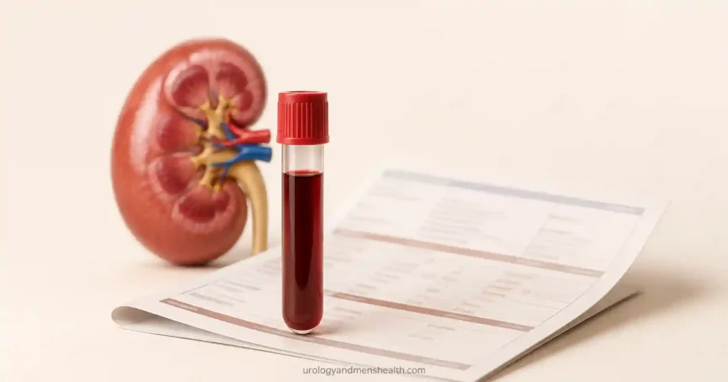
Protein in urine is one of the most common abnormal findings on a routine urinalysis, and one of the most badly explained. A man comes in holding a printout with a plus sign next to “protein,” having spent three days reading that it means kidney failure. In clinic, the majority of those results turn out to be transient — the product of a hot day, a gym session, or a fever the week before. But a meaningful minority are the earliest warning a kidney gives before anything else on the blood panel moves. The dipstick cannot tell those two groups apart, and neither can a single test of any kind. What separates them is repetition, the right follow-up test, and what else is sitting in the same urine sample. This article walks through what the finding actually represents at the level of the kidney filter, which causes are harmless and which are not, how the measurement is properly done, and what to ask for next.
Key Takeaways
- One positive dipstick is not a diagnosis — proteinuria is only called persistent when it appears on two or three separate samples across at least three months.
- Normal kidneys leak under 150 mg of total protein and under 30 mg of albumin per day; the albumin-to-creatinine ratio on a first-morning sample, not the dipstick, is the test that decides whether that line has been crossed.
- Protein and blood together in the same sample points at the kidney filter rather than the bladder or prostate — and per AUA guidance that means a nephrology referral plus the urology workup, not one instead of the other.
- An albumin-to-creatinine ratio above 30 mg/g raises cardiovascular risk independently of kidney function, which is why the result matters even when your creatinine and eGFR still read normal.
What Protein in Urine Actually Means
Your kidneys push around 180 liters of plasma (about 48 US gallons) through their filters every day. Almost all of it is reabsorbed. What passes through first has to cross a three-layer barrier inside each glomerulus: a perforated capillary lining, a dense basement membrane, and a lattice of specialized cells called podocytes whose interlocking foot processes leave slits just wide enough for water, salts, and small waste molecules.
That barrier filters by two properties at once — size and electrical charge. Albumin, the most abundant protein in blood, is both bulky and negatively charged, and the barrier itself carries a negative charge that repels it. Think of it as a sieve that is also a magnet set to push away exactly the thing you most want to keep.
Healthy kidneys still lose a trace. Under 150 mg of total protein per day is normal, and most of that is not albumin at all but Tamm-Horsfall protein, secreted by the tubules themselves. Normal albumin loss is under 30 mg per day. When either number climbs, one of three things has gone wrong: the barrier is leaking, the tubules have stopped reclaiming what slips through, or the blood is carrying so much of an abnormal protein that even an intact filter cannot hold it back. For the wider picture of how kidney and cardiovascular problems feed each other, our Blood Pressure & Kidney Hub collects the related reading in one place.
Causes of Protein in Urine: Harmless, Temporary, and Serious
The single most useful question is not what caused the protein but whether it is still there. Sort the causes into three buckets and the workup organizes itself.
Transient and functional — resolves without treatment
Fever, a hard workout, dehydration, cold exposure, and acute illness all raise urinary protein for hours to days. The mechanism is haemodynamic rather than structural — blood flow through the glomerulus shifts, the filter is briefly stressed, and it recovers. Decompensated heart failure does the same thing for longer. None of this leaves damage behind. It is also the reason a dipstick taken the morning after a marathon is close to worthless.
Orthostatic proteinuria belongs here too. Some men under 30 lose protein when upright but none in the first urine passed after lying flat all night. It is benign, it usually fades with age, and it is confirmed by comparing a first-morning sample with a daytime one. If the first-morning sample is clean, the case is closed.
Glomerular — the filter is leaking
This is the bucket that matters. Diabetes and long-standing high blood pressure are the two largest contributors worldwide, and both damage the barrier slowly enough that albumin appears years before creatinine moves. Beyond those sit the primary glomerular diseases — IgA nephropathy, membranous nephropathy, focal segmental glomerulosclerosis, minimal change disease — plus lupus, amyloidosis, and obesity-related glomerulopathy. These produce the largest protein losses, and the heaviest of them tip into nephrotic range: more than 3.5 g per day, with swelling, low blood albumin, and high cholesterol.
Tubular, overflow, and post-renal
- Tubular: the filter is fine but the tubules have stopped reabsorbing small proteins. Interstitial nephritis, long-term NSAID use, and heavy metal exposure are the usual culprits. Protein loss is modest and mostly non-albumin.
- Overflow: the blood carries an abnormal protein load the kidney was never designed for. Multiple myeloma light chains are the classic example — and notably, a standard dipstick reads albumin only, so it can be entirely negative while a man is spilling grams of light chain per day.
- Post-renal: infection, stones, inflammation, and catheters add protein to urine after it has left the kidney. Semen contamination of a sample does it too. Here the protein is a passenger, not a signal of filter damage.
In My Practice
A man in his forties was sent to me for foamy urine and a dipstick positive for both protein and blood, with a working assumption somewhere upstream that this was a prostate problem. His prostate was unremarkable for his age. What changed the direction of the consultation was the microscopy report, which nobody had opened: dysmorphic red cells and a red cell cast. That combination does not come from the bladder or the prostate. It comes from the glomerulus, and it turned a urology referral into a same-week nephrology referral and a biopsy that showed IgA nephropathy.
When protein and blood appear together, the AUA position is that you do both — refer for nephrological assessment and still complete the risk-based urologic evaluation, because a man can have glomerular disease and a bladder tumour at the same time.
How Protein in Urine Is Measured
Three tests exist and they are not interchangeable.
The dipstick is a colour-change reagent pad that responds almost entirely to albumin. It typically does not turn positive until albumin concentration reaches around 300 mg/L, which means it misses the moderately increased range where treatment decisions are actually made. It also reports concentration, not quantity — so a dehydrated man with concentrated urine reads positive on a normal protein load. Alkaline urine above pH 8, visible blood in the sample, and antiseptic contamination all produce false positives. The AUA makes the same argument about blood, stating that microhematuria should not be defined on dipstick alone [2]. The reasoning applies equally to protein.
The albumin-to-creatinine ratio (ACR) is the current standard, and it solves the concentration problem elegantly. Because urinary creatinine is excreted at a steady rate, dividing albumin by creatinine cancels out how dilute or concentrated the sample happens to be. One first-morning void gives a result that approximates a 24-hour collection. KDIGO 2024 guidance recommends testing at-risk people with both a urine albumin measurement and an eGFR, and repeating any incidental abnormal ACR rather than acting on it [1]. The categories:
- Under 30 mg/g (under 3 mg/mmol) — normal to mildly increased
- 30 to 300 mg/g (3 to 30 mg/mmol) — moderately increased; the range formerly labelled microalbuminuria
- Above 300 mg/g (above 30 mg/mmol) — severely increased
- Above roughly 2,200 mg/g — nephrotic range
The protein-to-creatinine ratio (PCR) and the 24-hour collection still have roles. PCR is requested when non-albumin protein is suspected — myeloma, tubular disease — because ACR would miss it entirely. The 24-hour jug remains the reference standard but is used sparingly now, since incomplete collection is common and introduces more error than it removes.
None of these are read alone. Serum creatinine and eGFR are ordered alongside, because the protein result and the function result answer different questions — a point covered in more detail in our guide to what a raised creatinine actually indicates.
Get the Blood Pressure & Kidney Protection Guide — what to test, what the numbers mean, and when to escalate
Enter your email below to receive Dr. Khalid’s complete Blood Pressure & Kidney Protection Guide as a free, printable PDF.
Why Proteinuria Predicts Kidney and Heart Damage
Kidney disease is not staged on function alone. It is staged on a grid — kidney function on one axis, albuminuria on the other — because the two carry independent prognostic weight [1]. Two men with an identical eGFR of 55 have materially different futures if one sits at 15 mg/g and the other at 400 mg/g. The albumin result is not a footnote to the function result. It is half the diagnosis.
There are two reasons for that. The first is that the glomerulus is a window onto blood vessels everywhere. A filtration barrier only leaks when its endothelium is damaged, and endothelium damaged in the kidney is rarely pristine in the coronary arteries. This is why albuminuria in the moderately increased range predicts cardiovascular events even in men whose kidney function never declines.
The second is that filtered protein is directly toxic to the tubules it passes through. Albumin arriving in the tubular lumen triggers an inflammatory response, and sustained inflammation drives interstitial scarring. Proteinuria is therefore both a marker of damage and a driver of it — which is what makes reducing it a treatment target in its own right, not merely a lab number to watch.
High blood pressure sits on both sides of this loop. Raised pressure transmitted into the glomerulus damages the barrier; the resulting protein loss accelerates scarring; scarring raises pressure further. Breaking that circuit early is the whole point of finding protein before symptoms exist, and the mechanics of it are set out in how high blood pressure damages your kidneys.
Where your ACR result places you on the CKD staging grid — stage by stageWhat to Do If Your Test Shows Protein in Urine
Work through this in order. Skipping step one is where most of the unnecessary anxiety and most of the unnecessary imaging comes from.
- Repeat it properly. Ask specifically for a first-morning urine albumin-to-creatinine ratio, not another dipstick. Do it at least 48 hours after any strenuous exercise, fever, or heavy dehydration. If it is abnormal, repeat again — persistence over three months is what defines chronic kidney disease, and a single result never does.
- Get the surrounding panel. Serum creatinine with eGFR, HbA1c or fasting glucose, a lipid profile, and urine microscopy. Ask directly whether the microscopy showed dysmorphic red cells or red cell casts — that answer changes the whole direction of the workup and is often not mentioned unless requested.
- Record your blood pressure properly. Two readings, morning and evening, for seven days, seated and rested. One clinic reading is not a blood pressure.
- Know the referral triggers. An ACR above 300 mg/g, protein alongside blood, an eGFR under 30, or a rapid fall in eGFR all warrant nephrology assessment rather than watchful waiting.
If proteinuria is confirmed as persistent, treatment aims at the number itself. An ACE inhibitor or ARB titrated to the maximum tolerated dose reduces protein loss beyond what its blood pressure effect alone would explain. SGLT2 inhibitors are now standard for albuminuric kidney disease with or without diabetes — dapagliflozin reduced kidney disease progression and cardiovascular death in the DAPA-CKD trial [3], and empagliflozin produced the same direction of benefit across a broader range of causes in EMPA-KIDNEY [4]. Sodium intake below 2,000 mg per day (about 5 g of salt, roughly one teaspoon) supports the drug effect. KDIGO guidance targets a systolic pressure below 120 mmHg using standardized office measurement in most adults with chronic kidney disease — a target that only means anything if the measurement technique matches the one the recommendation was built on [1].
To see where your own eGFR and albuminuria result sit before your next appointment, run both through our CKD Stage Calculator and take the printout with you.
Red Flags — Get Assessed Within Days, Not Months
Protein in urine is usually a slow-moving finding. These features are not, and they change the timeline:
- Swelling around the eyes on waking, or ankle and leg swelling that pits when pressed
- Frothy urine that persists on every void, especially with unexplained weight gain
- Protein together with visible or microscopic blood in the same sample
- Blood pressure above 180/110 mmHg on repeated readings
- A sharp drop in urine output, or a creatinine that has risen quickly between two recent tests
- Bone pain, unexplained anaemia, or fatigue alongside proteinuria — this combination raises the possibility of a plasma cell disorder that a standard dipstick cannot detect
Frequently Asked Questions
Does foamy urine always mean protein in urine?
No. Foam that clears within a minute is usually the force of the stream or residue in the bowl. Foam that persists after several minutes, appears on every void, and comes with ankle or eyelid swelling is more suggestive. Foam is a prompt to test, never a diagnosis — the only way to confirm protein in urine is a first-morning albumin-to-creatinine ratio.
Can dehydration or a hard workout cause a positive protein result?
Yes, and this is the most common reason a first dipstick misleads. Concentrated urine raises the protein reading without any kidney damage, and strenuous exercise, fever, or acute illness each produce transient proteinuria that clears within days. This is why one abnormal sample is never enough — repeating the test after 48 hours of normal hydration and no heavy exercise settles most cases.
What albumin-to-creatinine ratio should worry me?
Under 30 mg/g is normal. Between 30 and 300 mg/g is moderately increased, and even here cardiovascular risk rises measurably, which is where a formal assessment of your blood pressure and kidney damage risk earns its place. Above 300 mg/g is severely increased and warrants nephrology referral. Above roughly 2,200 mg/g is nephrotic range and needs urgent specialist assessment.
Can an enlarged prostate or a UTI cause protein in the urine?
They can, but only in small amounts and by a different mechanism. Infection, stones, and inflammation add white cells, blood, and protein to urine after it has left the kidney, so the protein is a passenger rather than evidence of filter damage. Treat the infection first, then retest. If protein persists once the urine is otherwise clear, the source is the kidney, not the prostate.
Will I need a kidney biopsy?
Most men with protein in urine never need one. A biopsy is considered when albuminuria is severe, when protein appears alongside dysmorphic red cells or red cell casts, when kidney function is falling quickly, or when the cause is still unclear after blood tests and imaging. Persistent moderate albuminuria with well-explained diabetes or hypertension is usually managed on treatment alone.
References
- Kidney Disease: Improving Global Outcomes (KDIGO) CKD Work Group. KDIGO 2024 Clinical Practice Guideline for the Evaluation and Management of Chronic Kidney Disease. Kidney International. 2024;105(4S):S117-S314. KDIGO
- American Urological Association / SUFU. Microhematuria: AUA/SUFU Guideline. 2020, amended 2025. AUA
- Heerspink HJL, Stefansson BV, Correa-Rotter R, et al. Dapagliflozin in patients with chronic kidney disease. New England Journal of Medicine. 2020;383(15):1436-1446. PubMed
- The EMPA-KIDNEY Collaborative Group. Empagliflozin in patients with chronic kidney disease. New England Journal of Medicine. 2023;388(2):117-127. PubMed
- National Institute of Diabetes and Digestive and Kidney Diseases. Chronic Kidney Disease: Tests and Diagnosis. NIDDK
Dr. Muhammad Khalid
MBBS · FCPS (Urology) · MCPS (General Surgery) · CHPE · CRSM · IMC #539472
Specialist urologist with 11+ years of clinical experience across tertiary teaching hospitals. Trained at Lady Reading Hospital and Khyber Teaching Hospital, Peshawar. Author or co-author of five peer-reviewed international publications, including in Cureus and the World Journal of Surgery. Procedural expertise: URS, PCNL, RIRS, TURP, TURBT, and major open urological surgery. Full profile →
This article is for educational purposes only and does not constitute medical advice. Always consult your physician or urologist for diagnosis and treatment decisions specific to your condition.