
Adrenal Incidentaloma Risk Stratifier
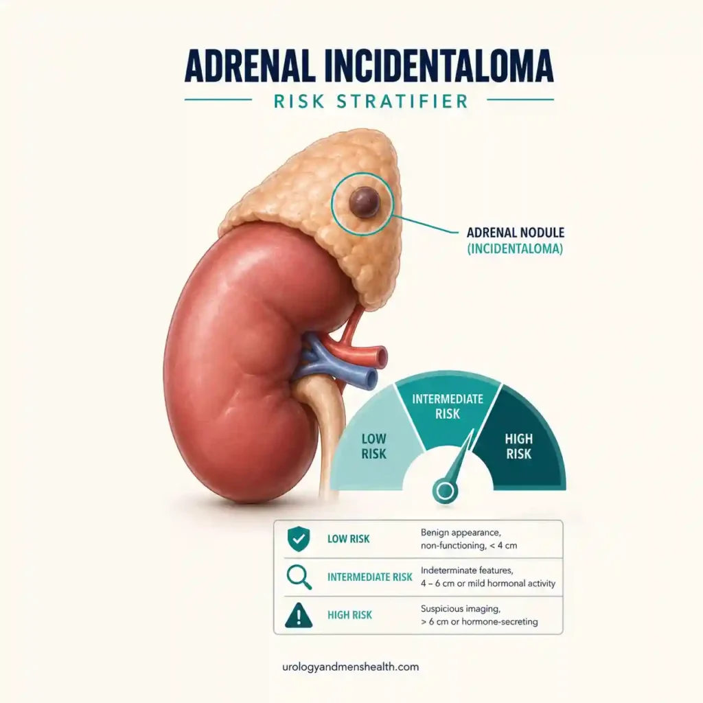
If a scan of your belly or chest turned up an adrenal nodule you were not looking for, this adrenal incidentaloma risk tool helps you read that finding the way a urologist would. Answer a few questions about the size, the density on your report, and your symptoms, and you will see whether it points to a routine check, a hormone work-up, or specialist review. Start with the Men's Wellness hub for the wider picture.

The Tool
Related Wellness Tools
Full Clinical Guide
In This Guide:
- Most adrenal incidentalomas are benign; a homogeneous mass at 10 HU or less on a plain CT is treated as benign whatever its size.
- Two things decide the plan: how the mass looks (size and density) and whether it makes a hormone.
- The standard first work-up is a 1-mg overnight dexamethasone test, plus metanephrines and an aldosterone-renin ratio when indicated.
- Size at or above 4 cm, an indeterminate look, or a cancer history means specialist review; recurring spells mean a pheochromocytoma must be excluded first.
What This Tool Measures
This tool estimates your adrenal incidentaloma risk – the chance that a nodule found by accident on your adrenal gland is something that needs acting on rather than simply watching. An adrenal incidentaloma is an adrenal lump picked up on a scan done for an unrelated reason, and it is common: roughly 3 to 4 percent of abdominal CT scans reveal one, and the figure climbs toward 10 percent in older adults [3]. The stratifier applies the risk logic of the two main clinical guidelines – the European ESE/ENSAT 2023 guideline and the American AACE/AAES guideline – which sort these masses by imaging appearance and by whether they overproduce a hormone [1][2]. The reassuring headline: about eight in ten turn out to be benign, non-functioning adenomas.
The Physiology Behind the Risk
Each adrenal gland is a small hormone factory sitting on top of a kidney. Two separate questions decide whether a nodule matters. First, what is it made of? Fat-rich (lipid-rich) tissue barely absorbs X-rays, so on a non-contrast CT it reads low – 10 Hounsfield units (HU) or less – and that reading alone marks it benign no matter how large it is [1]. Denser tissue (above 20 HU, or heterogeneous) does not absorb X-rays the same way and cannot be waved through. Second, is the factory running when it should be idle? A nodule can quietly overproduce cortisol, aldosterone, or adrenaline, and that can drive high blood pressure, a low potassium, or new diabetes – which is why the same nodule can matter more in a man whose blood pressure will not settle. If that describes you, the BP & Kidney Damage Risk tool is a useful companion check.
How to Read Your Result
The result routes you to one of three places rather than giving you a number. Lower concern means a small, lipid-rich mass with no worrying symptoms – your only job is confirming a one-time hormone screen. Needs a work-up means the imaging was not clearly benign, or something hints at a hormone problem, so ordinary blood tests come next. Specialist review means a feature crosses a line: a size at or above 4 cm, an indeterminate or heterogeneous look, a cancer history, or the spell pattern of a pheochromocytoma. The contrast is stark in clinic – a man with a 2 cm lipid-rich nodule and a man with a 5 cm heterogeneous one leave with completely different plans, even though both walked in with “a spot on my adrenal gland.” The 4 cm line exists because about nine in ten adrenal cancers are already larger than 4 cm when found [3][4].
Read: Secondary Hypertension – When High BP Has a Hidden CauseWhat to Do With Your Result
If you landed in lower concern, ask your doctor to record that a benign, lipid-rich mass needs no repeat scan, and complete the single hormone screen. If you need a work-up, the tests are specific: a 1-milligram overnight dexamethasone suppression test for cortisol (a morning level above 50 nmol/L, or 1.8 micrograms/dL, afterwards flags mild autonomous cortisol secretion), blood or urine metanephrines when the mass is above 10 HU, and an aldosterone-to-renin ratio if your blood pressure is high or your potassium runs low – with mineralocorticoid receptor blockers being one treatment if aldosterone is the culprit [1][2]. If you were routed to specialist review, that means dedicated adrenal imaging (washout CT or chemical-shift MRI) and an endocrine or urology opinion – and if you get pheochromocytoma-type spells, that tumor must be excluded before any needle goes near the mass. For the full picture of what the report words mean, see the companion cardiovascular tools once your blood pressure is being managed. If you are unsure about your result, the PDF report this tool generates gives you a ready-made framework to bring to your next appointment.
Read: Adrenal Incidentalomas – What the Scan Actually FoundIn My Practice
The commonest thing I undo is unnecessary repeat scanning. A man comes in having had the same small, clearly lipid-rich adenoma scanned three times in two years, because each new report says “stable adrenal nodule, recommend interval follow-up” and no one has stepped back to note it was benign from the very first plain CT.
Once a homogeneous mass has read 10 HU or below, the guideline is clear that it does not need chasing. Writing “benign adenoma, no further imaging” plainly in the notes saves the next doctor from restarting the cycle – and saves the patient a great deal of avoidable worry.
References
- Fassnacht M, Tsagarakis S, Terzolo M, et al. European Society of Endocrinology clinical practice guidelines on the management of adrenal incidentalomas, with ENSAT. Eur J Endocrinol. 2023;189(1):G1-G42. PubMed. ESE/ENSAT: <=10 HU benign; post-dexamethasone cortisol >50 nmol/L = mild autonomous cortisol secretion.
- Zeiger MA, Thompson GB, Duh QY, et al. AACE/AAES Medical Guidelines for the Management of Adrenal Incidentalomas. Endocr Pract. 2009;15(Suppl 1):1-20. PubMed. Screen all for cortisol excess; aldosterone if hypertensive; most masses >=4 cm resected.
- Kloos RT, et al. Radiologic Evaluation of Incidentally Discovered Adrenal Masses. Am Fam Physician. 2010;81(11):1361-1366. AAFP. Prevalence ~3-4%; ~90% of adrenocortical carcinomas are >4 cm at discovery.
- Sturgeon C, Shen WT, Clark OH, et al. Risk assessment in 457 adrenocortical carcinomas: how much does tumor size predict malignancy? J Am Coll Surg. 2006;202(3):423-430. PubMed. Malignancy likelihood rises with size above the 4 cm threshold.
Frequently Asked Questions
The scan found an “adrenal nodule” – does that mean cancer?
Almost always, no. About eight in ten adrenal incidentalomas are benign, non-functioning adenomas, and adrenocortical cancer is genuinely rare – on the order of one to two people per million each year. The words “nodule,” “lesion,” and “mass” describe a lump, not a diagnosis. What settles it is the density and size on your report, which is exactly what this tool reads. Our guide on what the scan actually found walks through the wording.
What does the Hounsfield unit (HU) number on my report mean?
Hounsfield units measure how dense tissue is on a CT scan. Fat-rich (lipid-rich) tissue reads low, and a homogeneous adrenal mass at 10 HU or less on a plain, non-contrast CT is treated as benign whatever its size – no repeat scan needed. Readings of 11 to 20 HU are indeterminate, and above 20 HU or “heterogeneous” needs further characterization. If your report gives no HU number, ask for a dedicated adrenal scan before anyone calls the mass benign.
Can an adrenal nodule be the reason my blood pressure is high?
Yes, sometimes. Some adrenal nodules overproduce aldosterone (causing high blood pressure, often with a low potassium) or, less often, adrenaline. That is why any hypertension is a reason to add an aldosterone-to-renin ratio to the work-up. If your blood pressure has been hard to control, it is worth checking whether the mass is the cause. You can gauge the wider impact with the BP & Kidney Damage Risk tool.
How accurate is this tool, and can I rely on it?
This tool applies the risk logic of the ESE/ENSAT 2023 and AACE/AAES guidelines to the information on your radiology report and your symptoms. It is a screening and education aid, not a diagnosis, and it cannot replace the radiologist’s reading or your doctor’s judgment. Some inputs, like the exact density and hormone results, come from tests only a clinician can order and interpret. Use the result to ask sharper questions, not as a final answer.
How do I use this result at my doctor’s appointment?
Tap “Download My Report” to generate a two-page PDF with your result, the interpretation, your suggested next steps, and a short list of questions to ask. Bring it, along with a copy of your radiology report, to your primary care doctor, urologist, or endocrinologist. It gives the visit a clear starting point – the size, the density, and which hormone tests still need doing – so nothing important is missed.
Dr. Muhammad Khalid
MBBS · FCPS (Urology) · MCPS (General Surgery) · CHPE · CRSM · IMC #539472
Specialist urologist with 11+ years of clinical experience across tertiary teaching hospitals. Trained at Lady Reading Hospital and Khyber Teaching Hospital, Peshawar. Author or co-author of five peer-reviewed international publications, including in Cureus and the World Journal of Surgery. Procedural expertise: URS, PCNL, RIRS, TURP, TURBT, and major open urological surgery. Full profile →
This article is for educational purposes only and does not constitute medical advice. Always consult your physician or urologist for diagnosis and treatment decisions specific to your condition.